
While we try to keep things accurate, this content is part of an ongoing experiment and may not always be reliable.
Please double-check important details — we’re not responsible for how the information is used.
Cosmetic Surgery
“Revolutionizing Eye-Tracking: New 3D Technology Paves Way for Next-Generation Applications”
By integrating powerful 3D imaging technology with advanced computation, researchers can capture gaze direction information from tens of thousands of surface points on the eye instead of about a dozen used by conventional eye-tracking methods. The technique could boost eye-tracking accuracy in a variety of fields ranging from the entertainment industry to medical research and industrial engineering.


Alternative Medicine
“Skin in a Syringe”: Breakthrough Technology Heals Burns without Scars
Scientists in Sweden have developed a groundbreaking “skin in a syringe” — a gel packed with live cells that can be applied directly to wounds or even 3D-printed into skin grafts. Designed to help the body build functional dermis rather than scar tissue, the innovation combines fibroblast cells on gelatin beads with a hyaluronic acid gel, held together using click chemistry. In a parallel advance, the team also created elastic hydrogel threads that can form tiny, fluid-carrying channels, paving the way for artificial tissues and organoid development.

Alternative Medicine
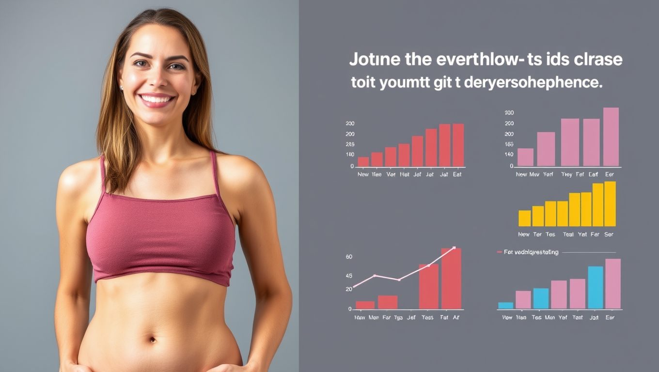
Patients Who Undergo Tummy Tuck Surgery Continue to Lose Weight Years Later, Study Finds
Patients who undergo tummy tuck surgery may be in for more than just cosmetic changes — a new study shows they often keep losing weight for years after the procedure. Researchers followed 188 patients and found consistent weight reduction up to five years later, especially in those with higher initial BMIs. Interestingly, lifestyle improvements, such as better diet and exercise habits, may play a key role in this surprising long-term effect. This could mean tummy tucks aren’t just sculpting bodies — they may be reshaping lives.
Cosmetic Surgery
“Microbes on Our Skin: The Hidden Heroes Against Sun Damage”
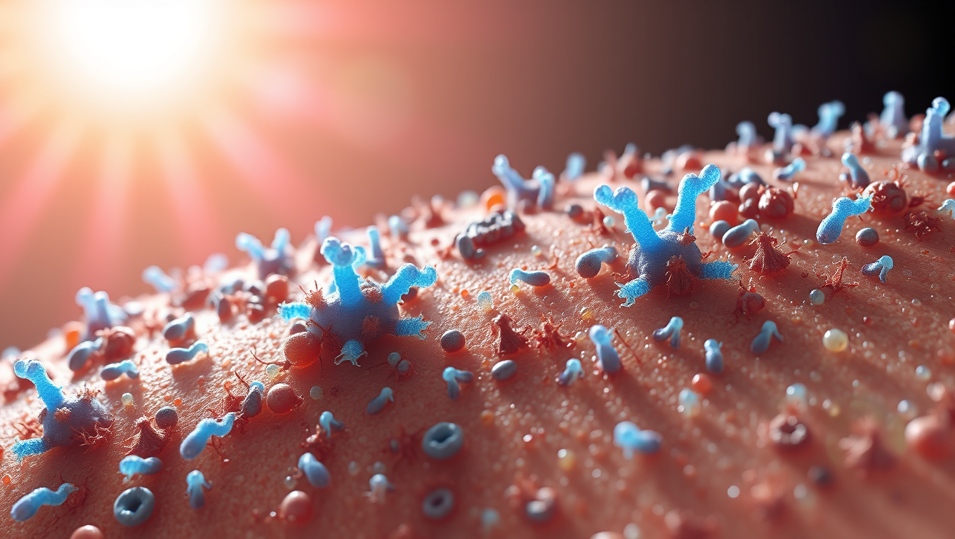
The skin microbiome plays an important role in health and disease. Researchers have now substantiated that certain skin bacteria can protect us from the sun’s ultraviolet (UV) radiation specifically by metabolizing cis-urocanic acid using an enzyme called urocanase. This enables the skin’s ability to fine-tune how it responds to UV radiation. The findings demonstrate the ability of the skin microbiome to remodel host immune functions.

A New Horizon for Vision: How Gold Nanoparticles May Restore People’s Sight

Revolutionizing Quantum Communication: Direct Connections Between Multiple Processors

Retiring Abroad Can Be Lonely Business
Harnessing Water Waves: A Breakthrough in Controlling Floating Objects

Household Electricity Three Times More Expensive Than Upcoming ‘Eco-Friendly’ Aviation E-Fuels, Study Reveals

“Unveiling Hidden Patterns: A New Twist on Interference Phenomena”
Reducing Falls Among Elderly Women with Polypharmacy through Exercise Intervention
-
 Detectors1 year ago
A New Horizon for Vision: How Gold Nanoparticles May Restore People’s Sight
-
Cancer1 year ago
Revolutionizing Quantum Communication: Direct Connections Between Multiple Processors
-
Earth & Climate1 year ago
Retiring Abroad Can Be Lonely Business
-
Albert Einstein1 year ago
Harnessing Water Waves: A Breakthrough in Controlling Floating Objects
-
Earth & Climate1 year ago
Household Electricity Three Times More Expensive Than Upcoming ‘Eco-Friendly’ Aviation E-Fuels, Study Reveals
-
Chemistry1 year ago
“Unveiling Hidden Patterns: A New Twist on Interference Phenomena”
-
Diseases and Conditions1 year ago
Reducing Falls Among Elderly Women with Polypharmacy through Exercise Intervention
-
Agriculture and Food1 year ago
“A Sustainable Solution: Researchers Create Hybrid Cheese with 25% Pea Protein”


