Connect with us

Animal Learning and Intelligence

Mapping the Mouse Brain: Unveiling the Secrets of Visual Perception and Connections

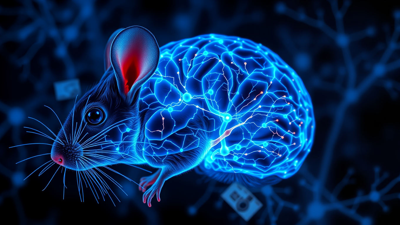
In a massive scientific effort, hundreds of researchers have helped to map the connections between hundreds of thousands of neurons in the mouse brain and then overlayed their firing patterns in response to visual stimuli. This breakthrough is a critical piece of foundational science to build toward understanding how our brains process visual information to reconstruct the images we see every day.
Avatar photo

Published

on

The scientists have made a groundbreaking discovery in understanding the intricacies of the mouse brain. In an unprecedented effort, hundreds of researchers worked together to map the connections between hundreds of thousands of neurons in the mouse brain and overlay their firing patterns in response to visual stimuli. This breakthrough is a crucial piece of foundational science that will help us comprehend how our brains process visual information, allowing us to reconstruct the images we see every day.

The human brain contains 86 billion neurons that make trillions of connections with each other through electrical firings. The complexity of its wiring diagram and the rapid movement of electrical signals across it in millisecond time frames hold the secrets of how our brain enables us to think, feel, and act. Although the current findings focus on a tiny fraction of the brain, they reveal the complex connections between cells and show how those connections are wired to produce functional responses.

To achieve this study, researchers presented video clips to mice genetically engineered for their neurons to emit light when they fire. The neuron firing patterns in areas associated with vision were optically recorded across a cubic millimeter – about the size of a grain of sand. Within this deceptively small amount of tissue lies remarkable complexity: four kilometers of axons, the processes that nerve cells use to communicate with each other, intertwined to make more than 524 million connections called synapses across more than 200,000 cells.

To map these connections, teams worked 12-hour shifts for 12 straight days to carefully cut and image ultra-thin slices of the brain tissue using electron microscopes. Reconstruction was the most challenging next step, as it required accurate stitching together almost 28,000 EM images to align the connections that cross the volume of brain tissue. This was followed by months of tracing the connections using deep learning algorithms, manual proofreading, and automated validation.

Deep learning predictive models were constructed and validated to explain visual information processing in the cortex. The sheer amount of data collected to create this tiny map comes out to 1.6 petabytes, roughly the equivalent of 22 years of continuous HD video.

These results come at a time when maps of neurons and their connections are increasingly revealing the mysteries of the brain. In 2023, research funded by the National Institutes of Health Brain Research Through Advancing Innovative Neurotechnologies Initiative produced the first complete cell atlas of the mouse brain, including the types and locations surveyed from more than 32 million cells. Last year, the NIH BRAIN Initiative “Flywire” project led to the complete mapping of the common fruit fly brain, demonstrating the unique value of mapping the whole brain in its entirety.

Funding for this project was provided through the Machine Intelligence from Cortical Networks Program of the Intelligence Advanced Research Projects Activity and the NIH BRAIN Initiative. The findings, published in a package of 10 papers published in the Nature family of journals, represent more than seven years of work performed by more than 150 scientists around the world.

Animal Learning and Intelligence

Harnessing the Power of Nature: How Black Tea and Berries Can Promote Healthy Aging

Higher intakes of black tea, berries, citrus fruits and apples could help to promote healthy aging, new research has found.

Avatar photo

Published

on

As we age, our bodies undergo various changes that can impact our physical and mental well-being. While some of these changes are inevitable, research suggests that incorporating certain foods into our diet can help promote healthy aging.

A recent study conducted by researchers from Edith Cowan University, Queen’s University Belfast, and Harvard T.H. Chan School of Public Health found that consuming higher intakes of black tea, berries, citrus fruits, and apples could lower the risk of key components of unhealthy aging, including frailty, impaired physical function, and poor mental health.

The study analyzed data from 62,743 women and 23,687 men over a period of 24 years. The findings revealed that women with the highest flavonoid intakes had a 15% lower risk of frailty, a 12% lower risk of impaired physical function, and a 12% lower risk of poor mental health compared to those with the lowest intakes.

While fewer associations were observed in men, higher flavonoid intake was still linked to a lower risk of poor mental health. The researchers suggested that regularly consuming flavonoid-rich foods could support healthier aging by reducing the risk of frailty, physical decline, and poor mental health.

The study’s lead author, Dr. Nicola Bondonno from Edith Cowan University, emphasized the importance of medical research in promoting healthy aging. “Our goal is not just to help people live longer but to ensure they stay healthy for as long as possible,” she said.

Professor Aedin Cassidy from Queen’s University Belfast added that flavonoids have been shown to reduce oxidative stress and inflammation, supporting blood vessel health, and even helping to maintain skeletal muscle mass – all of which are crucial for preventing frailty and maintaining physical function and mental health as we age.

The researchers noted that incorporating three servings of flavonoid-rich food into one’s diet could lead to a 6% to 11% lower risk across all three aging outcomes in females, and a 15% lower risk of poor mental health in males. This highlights the potential for simple dietary modifications to impact overall quality of life and contribute to the optimisation of healthy aging.

Overall, this study underscores the importance of incorporating flavonoid-rich foods into our diet to promote healthier aging. As Professor Eric Rimm from Harvard T.H. Chan School of Public Health noted, “These findings underscore the potential for simple dietary modifications to impact overall quality of life and contribute to the optimisation of healthy aging.”

Continue Reading

Animal Learning and Intelligence

Unlocking Orangutan Potential: How Zoo Life Boosts Exploration and Learning

A new study comparing wild and zoo-housed Sumatran orangutans reveals that life in a zoo significantly alters how orangutans interact with their environment. Researchers analyzed over 12,000 instances of daily exploratory object manipulation (EOM) — the active manipulation and visual inspection of objects associated with learning and problem-solving — across 51 orangutans aged 0.5 to 76 years. The findings show that orangutans living in zoos engage in more frequent, more diverse, and more complex exploration than their wild counterparts.

Avatar photo

Published

on

The lives of Sumatran orangutans living in zoos have been found to significantly alter how they interact with their environment, according to a recent study comparing wild and zoo-housed individuals. Researchers analyzed over 12,000 instances of daily exploratory object manipulation (EOM), revealing that orangutans in zoos engage in more frequent, diverse, and complex exploration than their wild counterparts.

“Our study shows that orangutans in zoos not only explore more but also explore differently,” said Isabelle Laumer, first author of the study. “What’s especially fascinating is that even when exploring the same kinds of objects, zoo-housed orangutans showed a richer repertoire of actions and were more likely to use tools or manipulate multiple objects at the same time.”

The study compared EOM behaviors in wild and zoo-housed orangutans across a wide age range. Data were collected from 33 wild individuals aged between six months and 76 years, as well as from 24 individuals in four zoos in Germany and Switzerland, with ages ranging from seven months to 49 years.

The results showed that zoo-housed orangutans explored objects more frequently than their wild counterparts, while there was no difference in exploration duration when individuals did explore. In contrast, wild orangutans primarily explored naturally occurring objects like plants, bark, and sticks, whereas zoo-housed orangutans engaged with a wider variety of enrichment items designed to encourage manipulation and cognitive engagement.

Importantly, the age at which orangutans first engaged in specific types of exploration was consistent across both settings, suggesting an innate developmental sequence. However, zoo orangutans continued to explore into adulthood, while wild individuals’ EOM declined sharply around weaning age (about 8 years old), likely due to the demands of survival in the wild.

In human infants, object exploration enables learning about physical properties such as texture and weight while stimulating cognitive and motor development – a pattern observed in many non-human animals as well. The heightened exploration may enhance cognitive flexibility and problem-solving skills in zoo-housed orangutans, as they interact with varied enrichment items and have more time and energy to devote to learning through exploration.

“These findings underscore how profoundly the environment influences animal behavior and cognitive development,” said Caroline Schuppli, senior author of the study. “And it also offers unique opportunities – by comparing wild and zoo-housed animals, we can better understand the full extent of a species’ cognitive potential.”

Continue Reading

Animal Learning and Intelligence

“Unlocking the Secrets of Animal Energy Consumption: A New Video-Based Method”

Strong methods do exist for measuring animal movement in the context of energy expenditure, but these are limited by the physical size of the equipment used. Now, in a paper published in the Journal of Experimental Biology, researchers from the Marine Biophysics Unit at the Okinawa Institute of Science and Technology (OIST), in collaboration with Professor Amatzia Genin from the Hebrew University of Jerusalem, describe an innovative method for measuring energy usage during movement with video and 3D-tracking via deep learning.

Avatar photo

Published

on

The amazing diversity of life on our planet is a testament to the multitude of biological solutions that have evolved to secure and maintain energy. However, despite its central role in biology, measuring energy consumption remains a challenging task. One major drain for many animals is movement, making it an ideal lens through which to estimate energy usage. While methods exist for measuring animal movement, they are often limited by the physical size of the equipment used.

In a groundbreaking study published in the Journal of Experimental Biology, researchers from the Marine Biophysics Unit at the Okinawa Institute of Science and Technology (OIST) have developed an innovative method for measuring energy usage during movement using video and 3D-tracking via deep learning. This new approach opens up the possibility of studying energy consumption in animals that were previously inaccessible due to the reliance on wearable equipment.

The current state-of-the-art method, Dynamic Body Acceleration (DBA), involves measuring oxygen consumption while an animal performs a specific behavior in a lab setting. However, this method has limitations when applied in the wild, where reliably measuring oxygen consumption is impossible. To overcome these challenges, researchers have used physical accelerometers that weigh at least ten times less than the animal, but this still rules out the study of many small species.

The OIST researchers’ solution to this problem is elegantly simple: they use two cameras to capture video footage of an animal’s behavior from multiple angles, reconstructing its movement in 3D space. A deep learning neural network is then trained on a few frames of the videos to track the position of body features such as eyes, allowing researchers to subsequently measure the movement-related acceleration.

This new video-based DBA method has opened up possibilities for studying energy consumption in animals that were previously inaccessible, potentially enabling many new research avenues into the breadth of life on our planet. For example, researchers can now investigate the energy expenditure during schooling of small fish, which has long remained mysterious. By accurately measuring energy usage during free-ranging animal behavior, scientists can gain a deeper understanding of the ecology and evolution of various species.

Continue Reading

Trending