Connect with us

Cell Biology

Unlocking the Secrets of Deadly Tropical Diseases: A New Target for Treatment

The efforts of a research team give hope for new treatment approaches for dangerous tropical diseases. The researchers have compiled a high-precision inventory of the membrane proteins of cell organelles of the African sleeping sickness pathogen.
Avatar photo

Published

on

The fight against deadly tropical diseases has taken a significant step forward with the discovery of a promising target for new treatments. Researchers from Bochum and Würzburg have made a groundbreaking find in their study on the African sleeping sickness pathogen Trypanosoma brucei, which also causes Chagas disease and leishmaniasis.

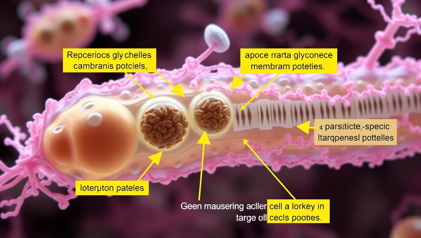
In a breakthrough study published in Cell Reports, the researchers compiled a high-precision inventory of the membrane proteins of the glycosomes, unique cell organelles essential for the survival of the parasites. “Some of these proteins contain components that are specific to parasites and differ significantly from those of the host cells,” explains Professor Ralf Erdmann.

The team’s success in identifying 28 glycosome membrane proteins with a high degree of reliability opens up new avenues for targeted treatment strategies against these poorly understood tropical diseases. A particular highlight was the discovery of TbPEX15, a membrane anchor for an essential protein import complex that differs significantly from its counterpart in humans.

This finding provides a valuable resource for biomedical research into glycosome biology and deepens our understanding of parasite biology. The researchers’ work gives hope for new treatment approaches for diseases that affect over 12 million people worldwide.

The study’s findings are a significant step forward in the fight against deadly tropical diseases, and further research could lead to the development of new therapies and treatments.

Biochemistry Research

A More Realistic Look at DNA in Action: Researchers Shed Light on the Mechanical Forces Behind Strand Separation

By creating a more true-to-life representation of DNA’s environment, researchers have discovered that strand separation may take more mechanical force than the field previously believed.

Avatar photo

Published

on

The way we study DNA has long been a topic of discussion among researchers. Traditionally, biochemistry labs isolate DNA within a water-based solution that allows scientists to manipulate it without interacting with other molecules. However, this approach can be misleading, as it doesn’t reflect the true environment of a living cell. In fact, the interior of a cell is “super crowded” with molecules, which can significantly impact the behavior of DNA.

Researchers at Northwestern University have taken a more realistic approach to studying DNA by creating an environment that mimics the conditions within a living cell. Led by Professor John Marko, the team used microscopic magnetic tweezers to separate DNA and then carefully attach strands of it to surfaces on one end, and tiny magnetic particles on the other. This allowed them to conduct high-tech imaging and investigate how different types of molecules interact with DNA.

The researchers found that strand separation, a crucial process for initiating replication or making repairs, may require more mechanical force than previously believed. They introduced three types of molecules to the solution holding DNA, mimicking proteins and investigating interactions among glycerol, ethylene glycol, and polyethylene glycol (each approximately the size of one DNA double helix, two or three nanometers).

“We wanted to have a wide variety of molecules where some cause dehydration, destabilizing DNA mechanically, and then others that stabilize DNA,” said Northwestern post-doctoral researcher Parth Desai. “It’s not exactly analogous to things found in cells, but you could imagine that other competing proteins in cells will have a similar effect.”
The team wrote a paper on their findings, which will be published on June 17 in the Biophysical Journal. Marko and Desai hope to run more experiments that incorporate multiple crowding agents and move closer to a true representation of a cell.

“If this affects DNA strand separation, all protein interactions with DNA are also going to be affected,” said Marko. “For example, the tendency for proteins to stick to specific sites on DNA and to control specific processes — this is also going to be altered by crowding.”

Their research has significant implications for understanding fundamental biochemical processes and may lead to new medical advances. The team hopes to study how interactions between enzymes and DNA are impacted by crowding in a living cell, which could have far-reaching consequences for our understanding of cellular biology.

This work was supported by the National Institutes of Health (grant R01-GM105847) and by subcontract to the University of Massachusetts Center for 3D Structure and Physics of the Genome (under NIH grant UM1-HG011536).

Continue Reading

Biology

“Disrupted Biological Clocks: The Hidden Dangers of Shift Work on Muscle Health and Aging”

Muscle cells contain their own circadian clocks and disrupting them with shift work can have a profound impact on aging, according to new research.

Avatar photo

Published

on

The article reveals that our biological clocks play a crucial role in maintaining muscle health and preventing accelerated aging, especially for shift workers. Research conducted by King’s College London has demonstrated how disrupting these internal timekeeping mechanisms can have a profound impact on our bodies.

The study focuses on the intrinsic muscle clock found within cells, which regulates protein turnover and is essential for muscle growth and function. At night, this clock activates the breakdown of defective proteins, replenishing muscles while the body rests. However, when this process is disrupted, it leads to muscle decline associated with aging, known as sarcopenia.

Using zebrafish in their research, the scientists found that impairing the muscle clock function resulted in premature aging, including reduced size, weight, and mobility. These findings are consistent with reports of shift workers experiencing similar health consequences.

The researchers also investigated protein turnover, a process critical for maintaining muscle mass. They discovered that during rest at night, the muscle clock regulates the degradation of defective muscle proteins, which accumulate throughout the day due to usage. This “nocturnal clearance” is essential for preserving muscle function, and its disruption may contribute to accelerated muscle decline.

The study’s lead author emphasizes the importance of understanding how circadian disruption affects shift workers’ health. With approximately four million shift workers in the UK, it is essential to develop strategies to improve their wellbeing. The researchers suggest that using circadian biology could lead to treatments aimed at preventing muscle decline in shift workers, paving the way for future therapies.

In conclusion, this study highlights the significance of biological clocks in maintaining muscle health and preventing accelerated aging, especially for shift workers. Further research is needed to develop effective strategies to mitigate these effects and improve the wellbeing of shift workers worldwide.

Continue Reading

Bacteria

Unlocking Efficiency: Researchers Reveal Secrets of Cell Division with Min Proteins

The Min protein system prevents abnormal cell division in bacteria, but is poorly understood. Researchers have uncovered how engineered e.coli bacteria control protein levels for maximum efficiency.

Avatar photo

Published

on

The Min protein system is a complex process that helps bacteria divide evenly and correctly. For decades, scientists have studied this system, but understanding how it works efficiently has been a challenge. Recently, researchers at the University of California San Diego (UCSD) made a groundbreaking discovery that sheds new light on the efficiency of cell division.

The UCSD team developed a way to control Min protein expression levels independently in E. coli cells. This allowed them to observe how different concentrations of Min proteins affect the oscillations between the poles of the cell. The results were surprising: despite varying concentrations, the oscillations remained stable across a wide range, with E. coli producing just the right amount of Min proteins.

This breakthrough is significant because it shows that the Min protein system can efficiently guide division to the correct location without relying on precise control over protein levels. This finding has far-reaching implications for our understanding of cellular organization and function.

The study was published in Nature Physics, a leading scientific journal, and was funded by the National Institutes of Health (NIH). The research team consisted of experts from both physics and chemistry/biochemistry departments at UCSD, highlighting the importance of interdisciplinary collaboration in advancing our knowledge of cellular biology.

Continue Reading

Trending