Connect with us
We’re experimenting with AI-generated content to help deliver information faster and more efficiently.
While we try to keep things accurate, this content is part of an ongoing experiment and may not always be reliable.
Please double-check important details — we’re not responsible for how the information is used.

Biochemistry

“Unlocking Hidden Risks: How MRI Scans Can Revolutionize the Detection of Life-Threatening Heart Disease”

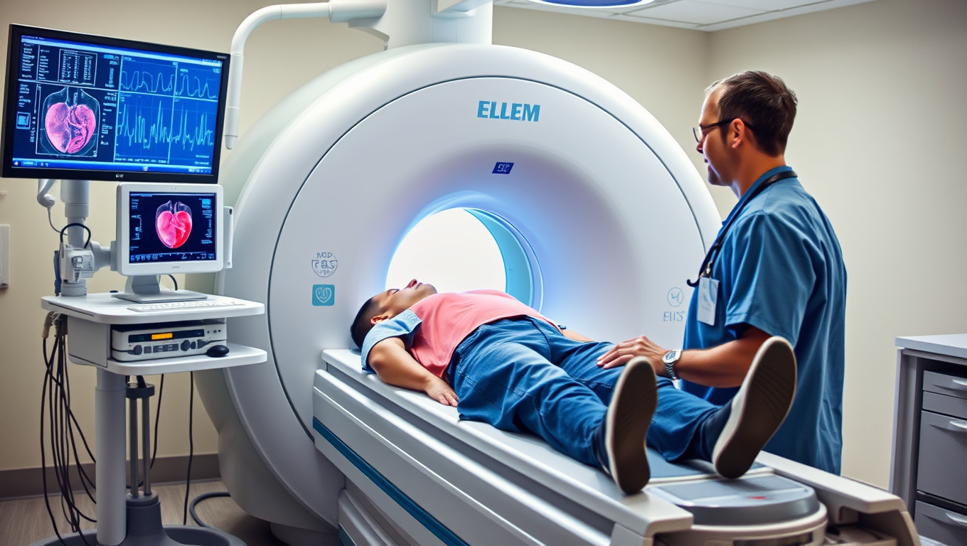
Magnetic resonance imaging (MRI) scans of the heart could help to detect a life-threatening heart disease and enable clinicians to better predict which patients are most at risk, according to a new study.
Avatar photo

Published

on

The article you provided is well-written, but I’ve made some changes to improve clarity, structure, and style for a general audience.

Lamin Heart Disease: A Hidden Killer

Lamin heart disease is a rare but potentially life-threatening condition that affects the heart’s ability to pump blood. It’s caused by a genetic mutation in the LMNA gene, which produces proteins essential for heart cell function. This condition often goes undiagnosed, affecting people in their 30s and 40s.

A new study led by researchers at University College London (UCL) has found that MRI scans can detect hidden signs of lamin heart disease, even when other tests show a healthy heart. The findings suggest that MRI could revolutionize the way we predict which patients are most at risk and inform decisions about life-changing treatments like heart transplants or implantable defibrillators.

Currently, risk estimates are based on electrocardiograms (ECGs), patient sex, genetics, symptoms, and basic heart function measurements by ultrasound. However, this tool underperforms for women and often recommends unnecessary devices that can be life-changing but not beneficial. The new study reveals the potential of cardiac MRI to improve disease risk predictions and become a standard part of managing lamin heart disease.

“Genetics alone cannot predict how this disease will progress,” said Dr. Gaby Captur, senior author of the study. “Two people with the same variant can have completely different outcomes.”

Lead author Dr. Cristian Topriceanu added, “Cardiac MRI picks up scarring of the heart tissue, inflammation, and signs the heart muscle is not working as well as it should among carriers of an LMNA mutation who do not have more overt signs of disease.”

These findings suggest a potential role for MRI in tracking disease progression and response to treatment. Gene therapies currently being trialled could tackle the cause of lamin heart disease, and MRI can identify people with early-stage disease displaying subtle abnormalities suggesting their disease is progressing.

The LMNA gene instructs the body to make proteins lamin A and C, critical to the structure and stability of heart cell nuclei. Mutations can lead to problems like dilated cardiomyopathy, life-threatening heart rhythms, and disrupted electrical signals regulating the heartbeat.

Close family members of individuals with lamin disease are screened for an LMNA mutation, but carriers usually only receive follow-up ECGs and echocardiograms. The new study suggests that cardiac MRI could become a standard tool in this process, providing earlier detection and more accurate risk assessments.

The research team analyzed MRI data from 187 people, finding heart damage, inflammation, and scarring central to lamin heart disease but not present in non-genetic dilated cardiomyopathy. Participants with a specific LMNA mutation showed worse heart functioning, highlighting the importance of cardiac MRI in understanding the mechanics of the heart.

This study has significant implications for the management of lamin heart disease, potentially revolutionizing the way we detect and treat this condition. As Dr. Captur noted, “Our findings show the potential of cardiac MRI to improve disease risk predictions and become a standard part of how we manage lamin heart disease.”

The study received funding from various organizations, including the British Heart Foundation, the National Institute of Health and Care Research (NIHR), Barts Charity, the Society for Cardiovascular Magnetic Resonance, and the NIHR UCLH Biomedical Research Centre.

In conclusion, the findings of this study highlight the importance of cardiac MRI in detecting hidden risks associated with lamin heart disease. This technology has the potential to revolutionize the way we predict which patients are most at risk and inform decisions about life-changing treatments like heart transplants or implantable defibrillators. As research continues to uncover the secrets of lamin heart disease, it’s clear that cardiac MRI will play a vital role in this journey.

Biochemistry

“Revolutionizing Medicine: A 100x Faster Path to Life-Saving Drugs with Metal Carbenes”

Using a clever combo of iron and radical chemistry, scientists have unlocked a safer, faster way to create carbenes molecular powerhouses key to modern medicine and materials. It s 100x more efficient than previous methods.

Avatar photo

Published

on

By

Chemists have made a groundbreaking breakthrough in developing a novel method to generate highly useful chemical building blocks by harnessing metal carbenes. This achievement is expected to revolutionize the synthesis of life-saving drugs and materials development.

Typically used in chemical reactions essential for drug synthesis, carbenes are short-lived, highly reactive carbon atoms. However, creating these carbenes has been a challenging task due to limited methods and hazardous procedures.

Researchers at The Ohio State University have now developed an approach that makes producing metal carbenes much easier and safer. According to David Nagib, co-author of the study and distinguished professor in arts and sciences, “Our goal all along was to determine if we could come up with new methods of accessing carbenes that others hadn’t found before.”

The team’s innovative method uses iron as a metal catalyst and combines it with chlorine-based molecules that easily generate free radicals. This combination works to form the carbene of their choice, including many that had never been made before.

These three-sided molecular fragments, known as cyclopropanes, are vital to the synthesis of medicines and agrichemicals due to their small size and unusual energy. The researchers’ work was inspired by looking for the best ways to create these shape, which is one of the most common found in medicines.

“Our lab is obsessed with trying to get the best methods for making cyclopropanes out there as soon as possible,” said Nagib. “We have the eye on the prize of inventing better tools to make better medicines, and along the way, we’ve solved a huge problem in the carbene world.”

The study was recently published in Science, and the team’s discovery is expected to become extremely impactful. By accessing a new way of creating and classifying carbenes, scientists can simplify and improve the current wasteful, multistep process of producing them.

For consumers, this method suggests that future drugs developed by this technology may be cheaper, more potent, faster-acting, and longer-lasting. The work could prevent shortages of important medicines like antibiotics and antidepressants, as well as drugs that treat heart disease, COVID, and HIV infections, said Nagib.

Additionally, the team would like to ensure that their transformational organic chemistry tool is accessible to both big and small research labs and drug manufacturers around the world. One way to guarantee this is by continuing to improve the current technique, said Nagib.

“Our team at Ohio State came together in the coolest, most collaborative way to develop this tool,” he said. “So we’re going to continue racing to show how many different types of catalysts it could work on and make all kinds of challenging and valuable molecules.”

Continue Reading

Biochemistry

Unveiling Molecular Motion: A Breakthrough in Synthetic Biology and Soft Matter Physics

Scientists have uncovered a previously unknown type of molecular motion inside DNA-based droplets: instead of spreading randomly, guest molecules advance in an organized wave. This surprising discovery opens the door to understanding how cells might organize internal processes without membranes. Using customizable DNA condensates as experimental models, the team showed how molecular waves emerge through precise DNA interactions. These insights could not only transform our grasp of cellular signaling but may even lay groundwork for treating neurodegenerative diseases by influencing how molecules behave inside aging cells.

Avatar photo

Published

on

Here’s the rewritten article:

In a groundbreaking discovery, researchers from Johannes Gutenberg University Mainz, the Max Planck Institute for Polymer Research, and the University of Texas at Austin have uncovered a form of molecular motion that defies conventional understanding. When guest molecules penetrate droplets of DNA polymers, they don’t diffuse haphazardly; instead, they propagate through them in a clearly-defined frontal wave.

“This is an effect we didn’t expect at all,” says Weixiang Chen, a leading researcher from the Department of Chemistry at JGU. The findings have been published in Nature Nanotechnology, and the implications are significant.

In contrast to traditional diffusion models, where molecules spread out randomly, the observed behavior of guest molecules in DNA droplets is structured and controlled. This takes the form of a wave of molecules or a mobile boundary, as explained by Professor Andreas Walther from JGU’s Department of Chemistry, who led the research project.

The researchers used thousands of individual strands of DNA to create droplets, known as biomolecular condensates. These structures can be precisely determined and have counterparts in biological cells, which employ similar condensates to arrange complex biochemical processes without membranes.

“Our synthetic droplets represent an excellent model system for simulating natural processes and improving our understanding of them,” emphasizes Chen.

The intriguing motion of guest molecules is attributed to the way that added DNA and the DNA present in the droplets combine on the basis of the key-and-lock principle. This results in swollen, dynamic states developing locally, driven by chemical binding, material conversion, and programmable DNA interactions.

The findings are not only fundamental to our understanding of soft matter physics but also relevant to improving our knowledge of cellular processes. “This might be one of the missing pieces of the puzzle that, once assembled, will reveal to us how cells regulate signals and organize processes on the molecular level,” states Walther.

This new insight could contribute to the treatment of neurodegenerative disorders, where proteins migrate from cell nuclei into the cytoplasm, forming condensates. As these age, they transform from a dynamic to a more stable state and build problematic fibrils. “It is quite conceivable that we may be able to find a way of influencing these aging processes with the aid of our new insights,” concludes Walther.

Continue Reading

Biochemistry

A Game-Changing mRNA Vaccine that’s More Effective and Less Costly to Develop

A new type of mRNA vaccine is more scalable and adaptable to continuously evolving viruses such as SARS-CoV-2 and H5N1, according to a new study.

Avatar photo

Published

on

A team of researchers from the University of Pittsburgh School of Public Health and Pennsylvania State University has made a groundbreaking discovery in the field of vaccine development. They have created a new type of mRNA vaccine that is not only more effective but also less costly to develop, making it a game-changer in the fight against infectious diseases.

The current mRNA vaccines, such as those used to prevent COVID-19, have two significant challenges: they require a high amount of mRNA to produce and are constantly evolving due to the changing nature of viruses like SARS-CoV-2 and H5N1. The researchers addressed these challenges by creating a proof-of-concept COVID-19 vaccine using what’s known as a “trans-amplifying” mRNA platform.

In this approach, the mRNA is separated into two fragments: the antigen sequence and the replicase sequence. The latter can be produced in advance, saving crucial time in the event of a new vaccine needing to be developed urgently and produced at scale. Additionally, the researchers analyzed the spike-protein sequences of all known variants of SARS-CoV-2 for commonalities, rendering what’s known as a “consensus spike protein” as the basis for the vaccine’s antigen.

The results are promising: in mice, the vaccine induced a robust immune response against many strains of SARS-CoV-2. This has the potential for more lasting immunity that would not require updating, because the vaccine has the potential to provide broad protection. Additionally, this format requires an mRNA dose 40 times less than conventional vaccines, so this new approach significantly reduces the overall cost of the vaccine.

The lessons learned from this study could inform more efficient vaccine development for other constantly evolving RNA viruses with pandemic potential, such as bird flu. The researchers hope to apply the principles of this lower-cost, broad-protection antigen design to pressing challenges like bird flu, making it a crucial step in preparing for future pandemics.

Continue Reading

Trending