While we try to keep things accurate, this content is part of an ongoing experiment and may not always be reliable.
Please double-check important details — we’re not responsible for how the information is used.
Disability
The Silent Threat: How Hearing Loss and Loneliness Fuel Memory Decline
A massive European study has uncovered a powerful connection between hearing loss, loneliness, and memory decline. Researchers at the University of Geneva found that older adults with hearing impairments who also feel lonely—regardless of actual social isolation—experience faster cognitive decline.


Alzheimer's
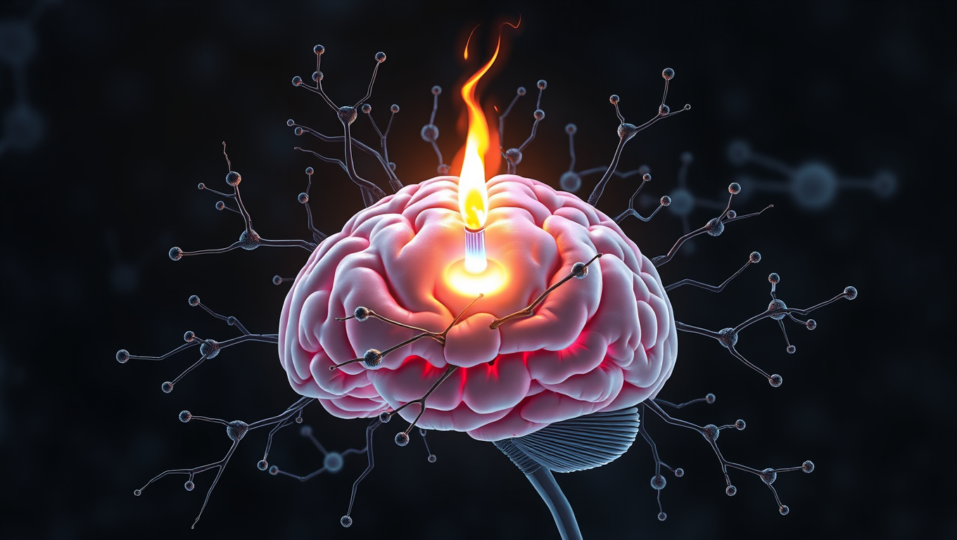
Scientists Unlock Secret to Reversing Memory Loss by Boosting Brain’s Energy Engines
Scientists have discovered a direct cause-and-effect link between faulty mitochondria and the memory loss seen in neurodegenerative diseases. By creating a novel tool to boost mitochondrial activity in mouse models, researchers restored memory performance, suggesting mitochondria could be a powerful new target for treatments. The findings not only shed light on the early drivers of brain cell degeneration but also open possibilities for slowing or even preventing diseases like Alzheimer’s.
Disability
“Lunar Trailblazer: A Small Satellite’s Quest to Map Water on the Moon Falls Short”
NASA’s Lunar Trailblazer, a mission designed to create high-resolution maps of water on the Moon, ended after losing contact with the spacecraft just one day after its February 26 launch. Despite extensive global efforts to reestablish communication, the small satellite’s misaligned solar arrays prevented its batteries from charging, leaving it powerless and drifting in a slow spin into deep space.

Alternative Medicine
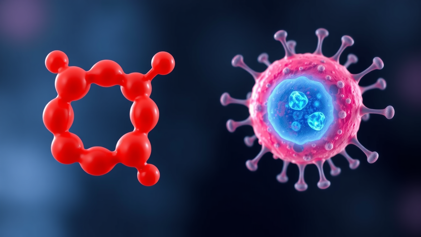
A Sweet Breakthrough: How a Sugar Molecule Could Help Treat Type 1 Diabetes
In a fascinating twist, Mayo Clinic researchers discovered that a sugar molecule cancer cells use to hide from the immune system might also protect insulin-producing beta cells in type 1 diabetes. By engineering these cells with the same sugar molecule—sialic acid—they prevented immune attacks in lab models. This approach could lead to better transplant options without broad immune suppression, offering hope for millions living with the autoimmune disease.

A New Horizon for Vision: How Gold Nanoparticles May Restore People’s Sight

Revolutionizing Quantum Communication: Direct Connections Between Multiple Processors

Retiring Abroad Can Be Lonely Business

“Unveiling Hidden Patterns: A New Twist on Interference Phenomena”
Harnessing Water Waves: A Breakthrough in Controlling Floating Objects

Household Electricity Three Times More Expensive Than Upcoming ‘Eco-Friendly’ Aviation E-Fuels, Study Reveals
Reducing Falls Among Elderly Women with Polypharmacy through Exercise Intervention
-
 Detectors1 year ago
A New Horizon for Vision: How Gold Nanoparticles May Restore People’s Sight
-
Cancer1 year ago
Revolutionizing Quantum Communication: Direct Connections Between Multiple Processors
-
Earth & Climate1 year ago
Retiring Abroad Can Be Lonely Business
-
Chemistry1 year ago
“Unveiling Hidden Patterns: A New Twist on Interference Phenomena”
-
Albert Einstein1 year ago
Harnessing Water Waves: A Breakthrough in Controlling Floating Objects
-
Earth & Climate1 year ago
Household Electricity Three Times More Expensive Than Upcoming ‘Eco-Friendly’ Aviation E-Fuels, Study Reveals
-
Diseases and Conditions1 year ago
Reducing Falls Among Elderly Women with Polypharmacy through Exercise Intervention
-
Acid Rain1 year ago
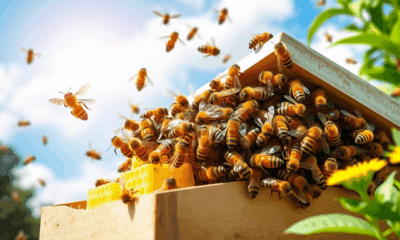
“Revolutionizing Honey Bee Survival: A New Pollen-Replacing Food Source Brings Hope for the Future”