Connect with us
We’re experimenting with AI-generated content to help deliver information faster and more efficiently.
While we try to keep things accurate, this content is part of an ongoing experiment and may not always be reliable.
Please double-check important details — we’re not responsible for how the information is used.

Autism

“Finding Balance: How Two Competing Nerve Proteins May Help Deter Autism Symptoms in Mice”

In mice, autism symptoms arise when a certain pair of competing nerve proteins falls out of equilibrium, according to a new study.
Avatar photo

Published

on

Autism Spectrum Disorder (ASD) affects approximately 1% of the global population, manifesting in a range of complex social and cognitive symptoms. While research has identified several genetic factors linked to ASD, particularly those related to neuron activity, the exact mechanisms behind these associations remain unclear. A recent study published in PLOS Biology sheds light on this mystery by examining the activities of two neuronal proteins suspected to be connected to ASD.

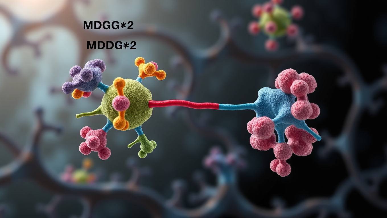
Researchers at Wenzhou Medical University and Xiamen University studied mice with reduced levels of MDGA2, a protein involved in transmitting nerve signals. Certain mutations in the MDGA2 gene have been identified in individuals with ASD. The experimental results showed that these mice exhibited ASD-like symptoms, including repetitive grooming and altered social behavior. Furthermore, they displayed increased activity in specific nerve synapses and elevated levels of BDNF, another neuronal protein linked to ASD.

Notably, when treated with an artificial peptide mimicking MDGA2, which inhibited BDNF/TrkB activity, the symptoms in these mice lessened. Based on this finding combined with previous research, the authors propose that MDGA2 and BDNF maintain a natural balance by competing for TrkB protein binding sites. This balance is essential for regulating neuron activity related to ASD. Disruptions to this system may lead to aberrant changes in excitatory neuronal activity.

This novel understanding of the MDGA2-BDNF-TrkB signaling pathway has significant implications for potential therapeutic treatments. Further investigation will be required to fully comprehend the functions of this system and its relationship to ASD symptoms. As Yun-wu Zhang notes, “Mutations in the MDGA2 gene cause autism spectrum disorders (ASD), but the underlying mechanism is elusive.” This study takes a crucial step towards illuminating this mystery, offering hope for future treatments that may help alleviate ASD symptoms in individuals affected by this condition.

Autism

“Unlocking Personalized Parkinson’s Treatment: Breakthrough Brain Scan Reveals Why Drugs Don’t Always Work”

Researchers are using an advanced brain imaging method called MEG to understand why Parkinson’s drug levodopa doesn’t work equally well for everyone. By mapping patients’ brain signals before and after taking the drug, they discovered that it sometimes activates the wrong brain regions, dampening its helpful effects. This breakthrough could pave the way for personalized treatment strategies, ensuring patients receive medications that target the right areas of their brain more effectively.

Avatar photo

Published

on

A groundbreaking study by Simon Fraser University researchers has shed new light on why Parkinson’s disease medications don’t always work as intended. By using a novel approach to brain imaging, the team found that the main drug used in dopamine replacement therapy – levodopa – can have unintended “off-target” effects in some patients.

Parkinson’s is the second most prevalent neurodegenerative disorder worldwide and affects millions of people globally. While levodopa is often effective in improving symptoms for many patients, it doesn’t work as well for everyone. To better understand why this is the case, researchers used magnetoencephalography (MEG) technology to study how the drug affects brain signals.

“We can see how levodopa activates certain parts of the brain in a patient,” said Alex Wiesman, assistant professor in biomedical physiology and kinesiology at SFU. “This information can help inform a more personalized approach to treatment.”

The study was a collaboration with researchers at Karolinska Institute in Sweden, who collected data from 17 patients with Parkinson’s disease using MEG technology. This advanced non-invasive technique measures the magnetic fields produced by the brain’s electrical signals.

Researchers mapped participants’ brain signals before and after taking the drug to see how it impacted brain activity. The results showed that some patients experienced “off-target” effects, which got in the way of the helpful effects of levodopa.

“We found that those people who showed ‘off target’ effects are still being helped by the drug, but not to the same extent as others,” Wiesman said.

The study’s findings have significant implications for personalized medicine. By understanding how individual patients respond to levodopa, clinicians may be able to adjust dosages or try different medications to improve treatment outcomes.

“This might be really helpful for tracking individualized responses to these types of drugs and helping with prescribing and therapeutics,” Wiesman said.

The new type of brain imaging analysis developed by the researchers is not only for studying Parkinson’s disease; any medications that affect brain signaling can be studied using this method. SFU’s ImageTech Lab, at the Surrey Memorial Hospital, is home to the only MEG in western Canada.

“Our next step is to take our new approach and apply it to a larger patient group,” Wiesman said. “We also need to translate this research to more accessible brain imaging methods, like electroencephalogram (EEG). Ultimately, we want to make sure this technology is useful for a diverse population and more widely accessible to patients with Parkinson’s disease.”

Continue Reading

Autism

Unpacking the Gene That Hijacks Fear: How PTEN Rewires the Brain’s Anxiety Circuit

Deleting a gene called PTEN in certain brain cells disrupts the brain’s fear circuitry and triggers anxiety-like behavior in mice — key traits seen in autism. Researchers mapped how this genetic tweak throws off the brain’s delicate balance of excitation and inhibition in the amygdala, offering deep insights into how one gene can drive specific ASD symptoms.

Avatar photo

Published

on

The gene PTEN has emerged as one of the most significant autism risk genes. Variations in this gene are found in a significant proportion of people with autism who also exhibit brain overgrowth. Researchers at the Max Planck Florida Institute for Neuroscience have discovered how loss of this gene rewires circuits and alters behavior, leading to increased fear learning and anxiety in mice – core traits seen in ASD.

PTEN has been linked to alterations in the function of inhibitory neurons in the development of ASD. The researchers focused on the changes in the central lateral amygdala driven by loss of PTEN in a critical neuronal population – somatostatin-expressing inhibitory neurons. They found that deleting PTEN specifically in these interneurons disrupted local inhibitory connectivity in the amygdala by roughly 50% and reduced the strength of the remaining inhibitory connections.

This diminished connectivity between inhibitory connections within the amygdala was contrasted by an increase in the strength of excitatory inputs received from the basolateral amygdala, a nearby brain region that relays emotionally-relevant sensory information to the amygdala. Behavioral analysis demonstrated that this imbalance in neural signaling was linked to heightened anxiety and increased fear learning, but not alterations in social behavior or repetitive behavior traits commonly observed in ASD.

The results confirm that PTEN loss in this specific cell type is sufficient to induce specific ASD-like behaviors and provide one of the most detailed maps to date of how local inhibitory networks in the amygdala are affected by genetic variations associated with neurological disorders. Importantly, the altered circuitry did not affect all ASD-relevant behaviors – social interactions remained largely intact – suggesting that PTEN-related anxiety and fear behaviors may stem from specific microcircuit changes.

By teasing out the local circuitry underlying specific traits, researchers hope to differentiate the roles of specific microcircuits within the umbrella of neurological disorders, which may one day help in developing targeted therapeutics for specific cognitive and behavioral characteristics. In future studies, they plan to evaluate these circuits in different genetic models to determine if these microcircuit alterations are convergent changes that underlie heightened fear and anxiety expression across diverse genetic profiles.

Continue Reading

Autism

CRISPR-edited stem cells hold key to understanding autism spectrum disorder

A team at Kobe University has created a game-changing resource for autism research: 63 mouse embryonic stem cell lines, each carrying a genetic mutation strongly associated with the disorder. By pairing classic stem cell manipulation with precise CRISPR gene editing, they ve built a standardized platform that mirrors autism-linked genetic conditions in mice. These models not only replicate autism-related traits but also expose key dysfunctions, like the brain s inability to clean up faulty proteins.

Avatar photo

Published

on

CRISPR technology has revolutionized genetics research, enabling scientists to edit genes with unprecedented precision. Recently, researchers at Kobe University developed a new method for modifying embryonic stem cells using CRISPR, creating a bank of 63 mouse embryonic stem cell lines containing the mutations most strongly associated with autism spectrum disorder (ASD). This breakthrough achievement has shed light on the hidden causes of ASD.

For decades, scientists have known that genetics play a significant role in the development of ASD. However, pinpointing the precise cause and mechanism remained elusive due to the lack of a standardized biological model for studying the effects of different mutations associated with the disorder. To address this challenge, Takumi Toru and his team at Kobe University embarked on a journey to create a reliable model by combining conventional manipulation techniques for mouse embryonic stem cells with CRISPR gene editing.

The new method proved highly efficient in making genetic variants of these cells, allowing the researchers to produce 63 mouse embryonic stem cell lines containing the mutations most strongly associated with ASD. These cell lines were further developed into various cell types and tissues, even generating adult mice with their genetic variations. The analysis of these cell lines revealed that autism-causing mutations often result in neurons being unable to eliminate misshapen proteins.

This finding is particularly interesting since the local production of proteins is a unique feature in neurons, and a lack of quality control of these proteins may be a causal factor of neuronal defects in ASD. Takumi expects that this achievement will be an invaluable resource for researchers studying autism and searching for drug targets. Moreover, the genetic variants studied are also implicated in other neuropsychiatric disorders such as schizophrenia and bipolar disorder, making this library potentially useful for studying these conditions as well.

This research was funded by various organizations, including the Japan Society for the Promotion of Science and the National Center of Neurology and Psychiatry. The study demonstrates the potential of CRISPR technology to reveal the hidden causes of complex diseases like ASD, paving the way for future discoveries and treatments.

Continue Reading

Trending