Connect with us
We’re experimenting with AI-generated content to help deliver information faster and more efficiently.
While we try to keep things accurate, this content is part of an ongoing experiment and may not always be reliable.
Please double-check important details — we’re not responsible for how the information is used.

Biology

From Dinosaurs to Birds: Unlocking the Secrets of Feather Formation

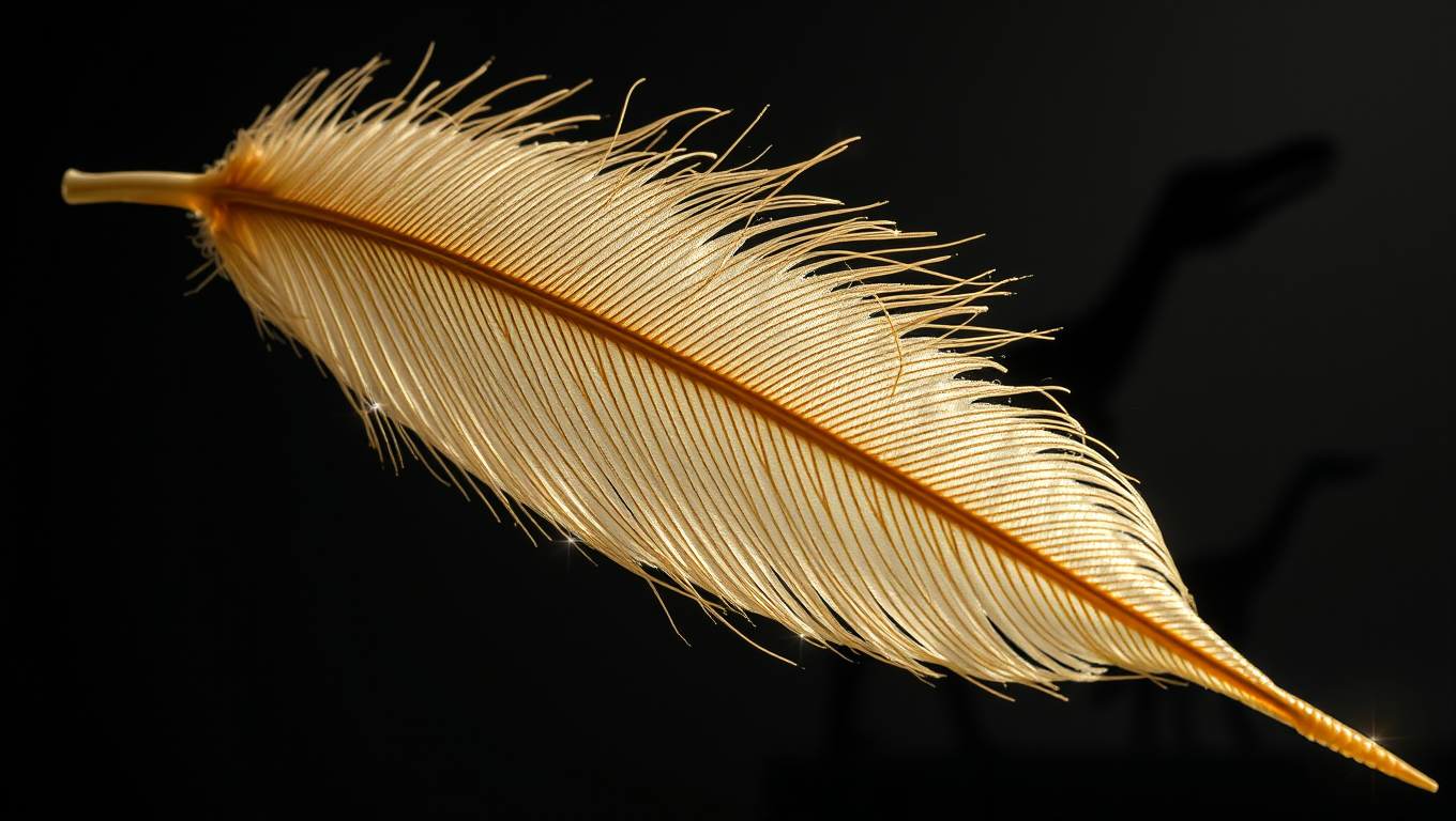
Feathers, essential for thermoregulation, flight, and communication in birds, originate from simple appendages known as proto-feathers, which were present in certain dinosaurs.By studying embryonic development of the chicken, researchers from the University of Geneva (UNIGE) have uncovered a key role of a molecular signalling pathway (the Shh pathway) in their formation. This research provides new insights into the morphogenetic mechanisms that led to feather diversification throughout evolution.
Avatar photo

Published

on

From Dinosaurs to Birds: Unlocking the Secrets of Feather Formation

The majestic plumage of birds has long fascinated scientists and the general public alike. However, the evolutionary journey that led to the development of feathers remains shrouded in mystery. A recent study by researchers from the University of Geneva has shed new light on this enigmatic process, revealing a crucial role played by molecular signaling pathways in feather formation.

Feathers are among the most complex cutaneous appendages in the animal kingdom. Their evolutionary origin has been extensively debated, with paleontological discoveries and developmental biology studies suggesting that feathers evolved from simple structures known as proto-feathers. These primitive filaments emerged around 200 million years ago in certain dinosaurs. Paleontologists continue to discuss the possibility of their even earlier presence in the common ancestor of dinosaurs and pterosaurs.

Proto-feathers are characterized by their simplicity, lacking barbs, barbules, and a follicle – an invagination at their base. The emergence of proto-feathers likely marked the first key step in feather evolution, providing thermal insulation and ornamentation before being progressively modified under natural selection to give rise to more complex structures enabling flight.

Researchers from Michel Milinkovitch’s laboratory at the University of Geneva have been studying the role of molecular signaling pathways, such as the Sonic Hedgehog (Shh) pathway, in the embryonic development of scales, hair, and feathers in modern vertebrates. In a previous study, they stimulated the Shh pathway by injecting an activating molecule into chicken embryos, observing the complete transformation of scales into feathers on the bird’s feet.

Building upon this knowledge, Rory Cooper and Michel Milinkovitch explored what happens when the Shh signaling pathway is inhibited. By injecting a molecule that blocks the Shh pathway on the 9th day of embryonic development – just before feather buds appear on the wings – they observed the formation of unbranched and non-invaginated buds, resembling the putative early stages of proto-feathers.

However, from the 14th day of embryonic development, feather morphogenesis partially recovered. The chicks hatched with patches of naked skin, but dormant subcutaneous follicles were autonomously reactivated, eventually producing chickens with normal plumage.

“Our experiments show that while a transient disturbance in the development of foot scales can permanently turn them into feathers, it is much harder to permanently disrupt feather development itself,” concludes Michel Milinkovitch. “Clearly, over the course of evolution, the network of interacting genes has become extremely robust, ensuring the proper development of feathers even under substantial genetic or environmental perturbations.”

The study provides new insights into the morphogenetic mechanisms that led to feather diversification throughout evolution. The findings highlight the complexity and resilience of biological systems, emphasizing the need for further research to understand how genetic interactions evolve to allow for the emergence of morphological novelties like proto-feathers.

Biochemistry Research

The Whispering Womb: Uncovering the Secret Language of Embryonic Cells

Scientists found that embryonic skin cells “whisper” through faint mechanical tugs, using the same force-sensing proteins that make our ears ultrasensitive. By syncing these micro-movements, the cells choreograph the embryo’s shape, a dance captured with AI-powered imaging and computer models. Blocking the cells’ ability to feel the whispers stalls development, hinting that life’s first instructions are mechanical. The discovery suggests hearing hijacked an ancient force-sensing toolkit originally meant for building bodies.

Avatar photo

Published

on

The human body begins as a single cell that multiplies and differentiates into thousands of specialized cells. Researchers at the Göttingen Campus Institute for Dynamics of Biological Networks (CIDBN) and the Max Planck Institute have made a groundbreaking discovery: embryonic cells “listen” to each other through molecular mechanisms previously known only from hearing.

Using an interdisciplinary approach combining developmental genetics, brain research, hearing research, and theoretical physics, the researchers found that in thin layers of skin, cells register the movements of their neighboring cells and synchronize their own tiny movements with those of the others. This coordination allows groups of neighboring cells to pull together with greater force, making them highly sensitive and able to respond quickly and flexibly.

The researchers created computer models of tissue development, which showed that this “whispering” among neighboring cells leads to an intricate choreography of the entire tissue, protecting it from external forces. These findings were confirmed by video recordings of embryonic development and further experiments.

Dr. Matthias Häring, group leader at the CIDBN, explained that using AI methods and computer-assisted analysis allowed them to examine about a hundred times more cell pairs than was previously possible in this field, giving their results high accuracy.

The mechanisms revealed in embryonic development are also known to play a role in hearing, where hair cells convert sound waves into nerve signals. The ear is sensitive because of special proteins that convert mechanical forces into electrical currents. This discovery suggests that such sensors of force may have evolved from our single-celled ancestors, which emerged long before the origin of animal life.

Professor Fred Wolf, Director of the CIDBN, noted that future work should determine whether the original function of these cellular “nanomachines” was to perceive forces inside the body rather than perceiving the outside world. This phenomenon could provide insights into how force perception at a cellular level has evolved.

Continue Reading

Biochemistry Research

Unlocking the Secrets of Life: A Spontaneous Reaction that Could Have Started it All

Scientists have uncovered a surprising new way that urea—an essential building block for life—could have formed on the early Earth. Instead of requiring high temperatures or complex catalysts, this process occurs naturally on the surface of tiny water droplets like those in sea spray or fog. At this boundary between air and water, a unique chemical environment allows carbon dioxide and ammonia to combine and spontaneously produce urea, without any added energy. The finding offers a compelling clue in the mystery of life’s origins and hints that nature may have used simple, everyday phenomena to spark complex biological chemistry.

Avatar photo

Published

on

The discovery of a previously unknown reaction pathway for the formation of urea has shed new light on the origins of life. A research team led by Ruth Signorell, Professor of Physical Chemistry at ETH Zurich, has made this groundbreaking finding, which has been published in the journal Science.

Until now, the industrial production of urea required high pressures and temperatures or chemical catalysts. However, enzymes enable the same reaction to take place in humans and animals, removing toxic ammonia from the breakdown of proteins such as urea. As this simple molecule contains nitrogen as well as carbon and probably existed on the uninhabited Early Earth, many researchers view urea as a possible precursor for complex biomolecules.

Signorell’s team studied tiny water droplets, such as those found in sea spray and fine mist. The researchers observed that urea can form spontaneously from carbon dioxide (CO2) and ammonia (NH₃) in the surface layer of the droplets under ambient conditions. This remarkable reaction takes place without any external energy supply.

The physical interface between air and liquid creates a special chemical environment at the water surface that makes the spontaneous reaction possible. Chemical concentration gradients form in this area, which acts like a microscopic reactor. The pH gradient across the interfacial layer of the water droplets creates the required acidic environment, which opens unconventional pathways that would otherwise not take place in liquids.

The results suggest that this natural reaction could also have been possible in the atmosphere of the Early Earth — an atmosphere that was rich in CO2 and probably contained small traces of ammonia. In such environments, aqueous aerosols or fog droplets could have acted as natural reactors in which precursor molecules such as urea were formed.

In the long term, the direct reaction of CO2 and ammonia under ambient conditions could also have potential for the climate-friendly production of urea and downstream products. This study opens a new window into the early days of the Earth and provides valuable insights into processes that could be significant for evolution.

The discovery of this spontaneous reaction pathway has significant implications for our understanding of the origins of life. It suggests that seemingly mundane interfaces can become dynamic reaction spaces, and biological molecules may have a more common origin than was previously thought.

Continue Reading

Bacteria

Unveiling the Secrets of Pandoraea: How Lung Bacteria Forge Iron-Stealing Weapons to Survive

Researchers investigating the enigmatic and antibiotic-resistant Pandoraea bacteria have uncovered a surprising twist: these pathogens don’t just pose risks they also produce powerful natural compounds. By studying a newly discovered gene cluster called pan, scientists identified two novel molecules Pandorabactin A and B that allow the bacteria to steal iron from their environment, giving them a survival edge in iron-poor places like the human body. These molecules also sabotage rival bacteria by starving them of iron, potentially reshaping microbial communities in diseases like cystic fibrosis.

Avatar photo

Published

on

As scientists continue to unravel the mysteries of the human microbiome, a team of researchers has made a groundbreaking discovery about the lung bacteria Pandoraea. These microbes have long been associated with disease-causing properties, but new research reveals that they also possess remarkable survival strategies, including the ability to forge iron-stealing weapons to thrive in challenging environments.

At the Leibniz Institute for Natural Product Research and Infection Biology (Leibniz-HKI), researchers led by Elena Herzog have been studying Pandoraea bacteria, which are known to be pathogenic but also produce natural products with antibacterial effects. The team’s investigation has shed light on how these bacteria manage to survive in iron-poor environments within the human body.

Iron plays a vital role in living organisms, including bacteria, as it is essential for enzymes and the respiratory chain. However, in environments like the human body, where iron is scarce, microorganisms must adapt to compete for this essential resource. Pandoraea bacteria have developed a unique strategy by producing siderophores – small molecules that bind iron from their environment and transport it into the cell.

The researchers identified a previously unknown gene cluster called pan, which codes for a non-ribosomal peptide synthetase enzyme responsible for the production of siderophores. Through targeted inactivation of genes and advanced analytical techniques, they isolated two new natural products, Pandorabactin A and B, which can complex iron and play an important role in how Pandoraea strains survive.

Moreover, bioassays revealed that pandorabactins inhibit the growth of other bacteria by removing iron from these competitors. The researchers also analyzed sputum samples from cystic fibrosis patients, finding that the detection of the pan gene cluster correlates with changes in the lung microbiome. This suggests that pandorabactins could have a direct influence on microbial communities in diseased lungs.

While the study’s findings are still preliminary and not yet suitable for medical applications, they provide valuable insights into the survival strategies of Pandoraea bacteria and the complex competition for vital resources within the human body. As researchers continue to explore the intricacies of the microbiome, this discovery paves the way for further investigation and potentially innovative treatments in the future.

Continue Reading

Trending