
While we try to keep things accurate, this content is part of an ongoing experiment and may not always be reliable.
Please double-check important details — we’re not responsible for how the information is used.
Bone and Spine
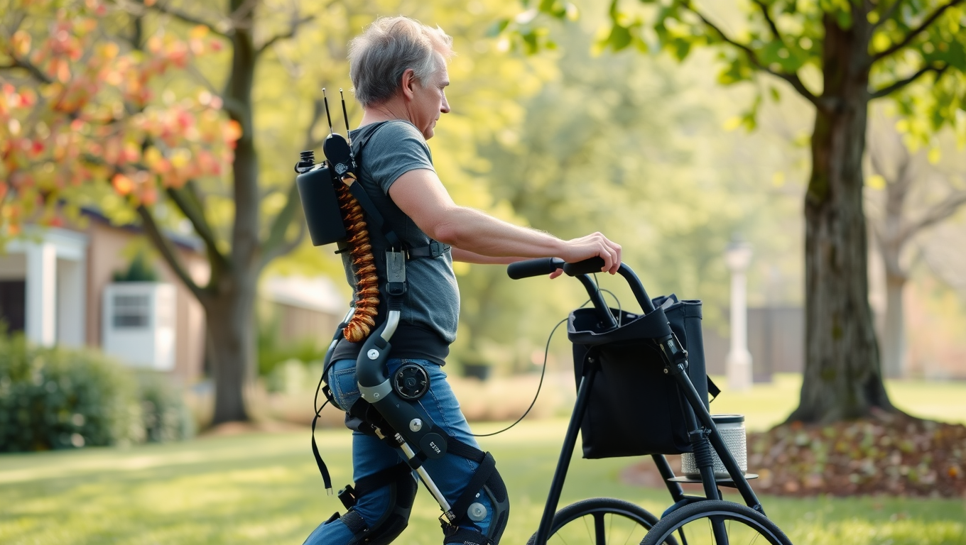
“Revolutionizing Rehabilitation: Robotics and Spinal Stimulation Restore Movement in Paralysis”
Scientists have developed an approach that combines rehabilitation robotics with spinal cord stimulation to restore movement in people with spinal cord injuries. The technology enhances rehabilitation and enables activities like cycling and walking outdoors.


Alternative Medicine
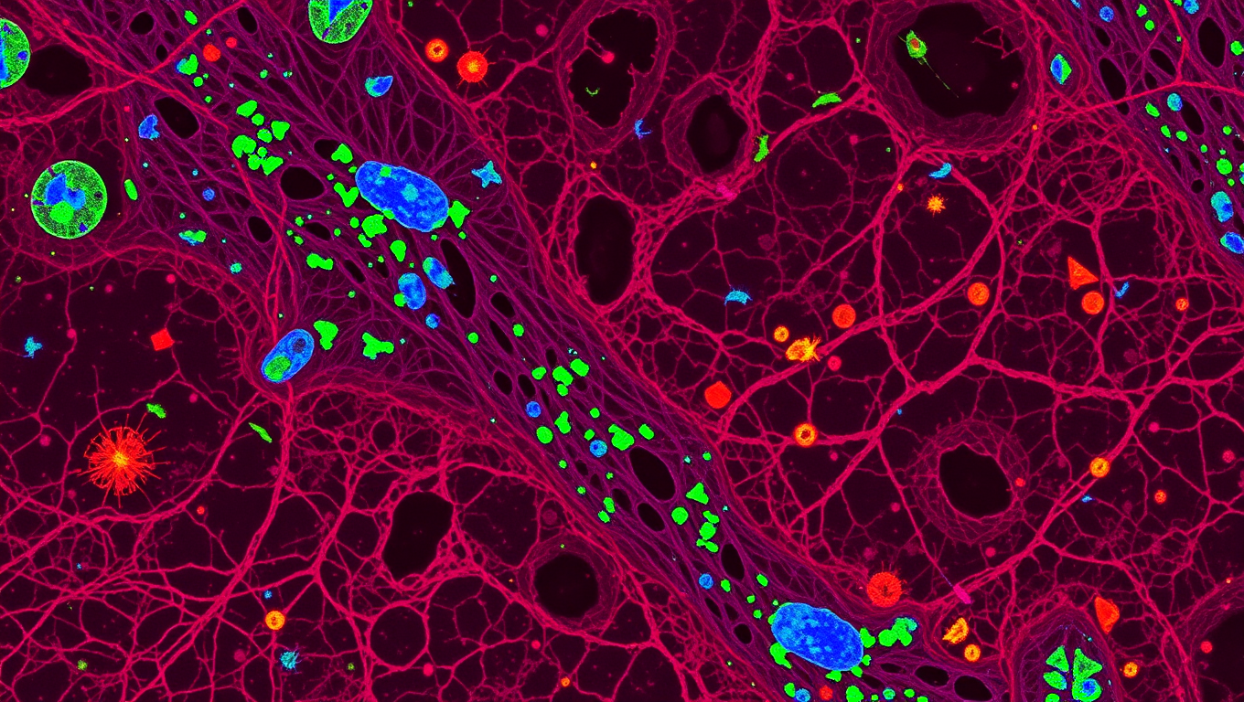
Breaking Barriers in Diabetic Wound Healing: A Revolutionary “Smart” Gel Accelerates Blood Flow and Restores Tissue Repair
A new gel-based treatment could change the way diabetic wounds heal. By combining tiny healing messengers called vesicles with a special hydrogel, scientists have created a dressing that restores blood flow and helps wounds close much faster. In tests, the treatment healed diabetic wounds far quicker than normal, while also encouraging the growth of new blood vessels. Researchers believe this innovation could one day help millions of people with slow-healing wounds caused by diabetes and possibly other conditions.

Bone and Spine
The Quiet Side Effect of Semaglutide: How Losing Muscle Can Affect Weight Loss Success
Semaglutide, a popular anti-obesity drug, may come with a hidden cost: significant muscle loss, especially in women and older adults. A small study found that up to 40% of weight loss from semaglutide comes from lean body mass. Alarmingly, those who consumed less protein saw even more muscle loss—potentially undermining improvements in blood sugar control.

Bone and Spine
Unlocking New Insights into Bone Marrow: Scientists Develop Revolutionary Imaging Technique
A new bone marrow imaging technique could change treatment for cancer, autoimmune disease and musculoskeletal disorders.

A New Horizon for Vision: How Gold Nanoparticles May Restore People’s Sight

Revolutionizing Quantum Communication: Direct Connections Between Multiple Processors

Retiring Abroad Can Be Lonely Business
Harnessing Water Waves: A Breakthrough in Controlling Floating Objects

“Unveiling Hidden Patterns: A New Twist on Interference Phenomena”

Household Electricity Three Times More Expensive Than Upcoming ‘Eco-Friendly’ Aviation E-Fuels, Study Reveals
Reducing Falls Among Elderly Women with Polypharmacy through Exercise Intervention
-
 Detectors1 year ago
A New Horizon for Vision: How Gold Nanoparticles May Restore People’s Sight
-
Cancer1 year ago
Revolutionizing Quantum Communication: Direct Connections Between Multiple Processors
-
Earth & Climate1 year ago
Retiring Abroad Can Be Lonely Business
-
Albert Einstein1 year ago
Harnessing Water Waves: A Breakthrough in Controlling Floating Objects
-
Chemistry1 year ago
“Unveiling Hidden Patterns: A New Twist on Interference Phenomena”
-
Earth & Climate1 year ago
Household Electricity Three Times More Expensive Than Upcoming ‘Eco-Friendly’ Aviation E-Fuels, Study Reveals
-
Diseases and Conditions1 year ago
Reducing Falls Among Elderly Women with Polypharmacy through Exercise Intervention
-
Agriculture and Food1 year ago
“A Sustainable Solution: Researchers Create Hybrid Cheese with 25% Pea Protein”


