
While we try to keep things accurate, this content is part of an ongoing experiment and may not always be reliable.
Please double-check important details — we’re not responsible for how the information is used.
Autism
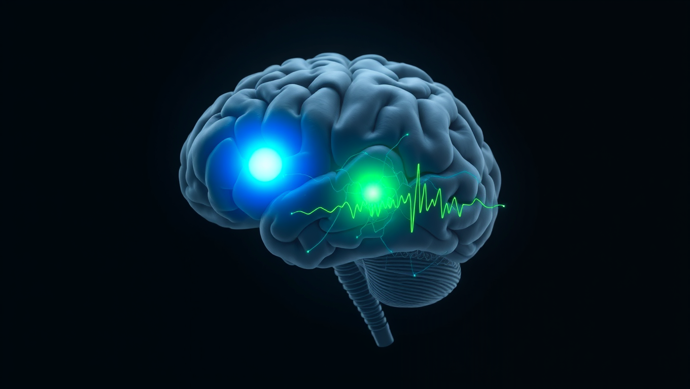
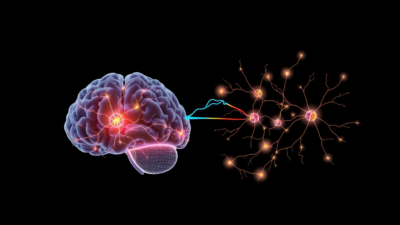
The Brain’s Hidden Patterns: Uncovering the Secret to Flexibility and Stability
A new study challenges a decades-old assumption in neuroscience by showing that the brain uses distinct transmission sites — not a shared site — to achieve different types of plasticity.


Autism
The Thalamic Feedback Loop: Unveiling the Brain’s Secret Pathway to Sensory Perception
Sometimes a gentle touch feels sharp and distinct, other times it fades into the background. This inconsistency isn’t just mood—it’s biology. Scientists found that the thalamus doesn’t just relay sensory signals—it fine-tunes how the brain responds to them, effectively changing what we feel. A hidden receptor in the cortex seems to prime neurons, making them more sensitive to touch.
Autism
The Hidden Power of Eye Contact: Unlocking Human Connection in Technology
A groundbreaking study from Flinders University reveals that it’s not just making eye contact that matters, but precisely when and how you do it. By studying interactions between humans and virtual partners, researchers discovered a powerful gaze sequence that makes people more likely to interpret a look as a call for help. Even more surprising: the same response pattern held true whether the “partner” was human or robot, offering insights into how our brains instinctively process social cues.

Autism
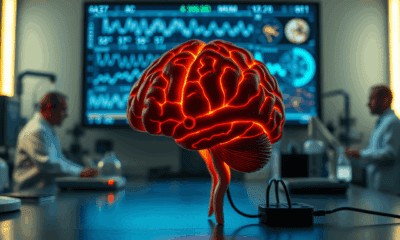
“Unlocking Personalized Parkinson’s Treatment: Breakthrough Brain Scan Reveals Why Drugs Don’t Always Work”
Researchers are using an advanced brain imaging method called MEG to understand why Parkinson’s drug levodopa doesn’t work equally well for everyone. By mapping patients’ brain signals before and after taking the drug, they discovered that it sometimes activates the wrong brain regions, dampening its helpful effects. This breakthrough could pave the way for personalized treatment strategies, ensuring patients receive medications that target the right areas of their brain more effectively.

A New Horizon for Vision: How Gold Nanoparticles May Restore People’s Sight

Retiring Abroad Can Be Lonely Business

Revolutionizing Quantum Communication: Direct Connections Between Multiple Processors
Harnessing Water Waves: A Breakthrough in Controlling Floating Objects

“Unveiling Hidden Patterns: A New Twist on Interference Phenomena”

Household Electricity Three Times More Expensive Than Upcoming ‘Eco-Friendly’ Aviation E-Fuels, Study Reveals

“A Sustainable Solution: Researchers Create Hybrid Cheese with 25% Pea Protein”
-
 Detectors11 months ago
A New Horizon for Vision: How Gold Nanoparticles May Restore People’s Sight
-
Earth & Climate12 months ago
Retiring Abroad Can Be Lonely Business
-
Cancer12 months ago
Revolutionizing Quantum Communication: Direct Connections Between Multiple Processors
-
Albert Einstein1 year ago
Harnessing Water Waves: A Breakthrough in Controlling Floating Objects
-
Chemistry12 months ago
“Unveiling Hidden Patterns: A New Twist on Interference Phenomena”
-
Earth & Climate12 months ago
Household Electricity Three Times More Expensive Than Upcoming ‘Eco-Friendly’ Aviation E-Fuels, Study Reveals
-
Agriculture and Food12 months ago
“A Sustainable Solution: Researchers Create Hybrid Cheese with 25% Pea Protein”
-
Diseases and Conditions1 year ago
Reducing Falls Among Elderly Women with Polypharmacy through Exercise Intervention


