Connect with us

Biochemistry Research

The Hidden Cost of Young Plant Disease Resistance

A new study reveals an evolutionary trade-off that young plants face to develop disease resistance.
Avatar photo

Published

on

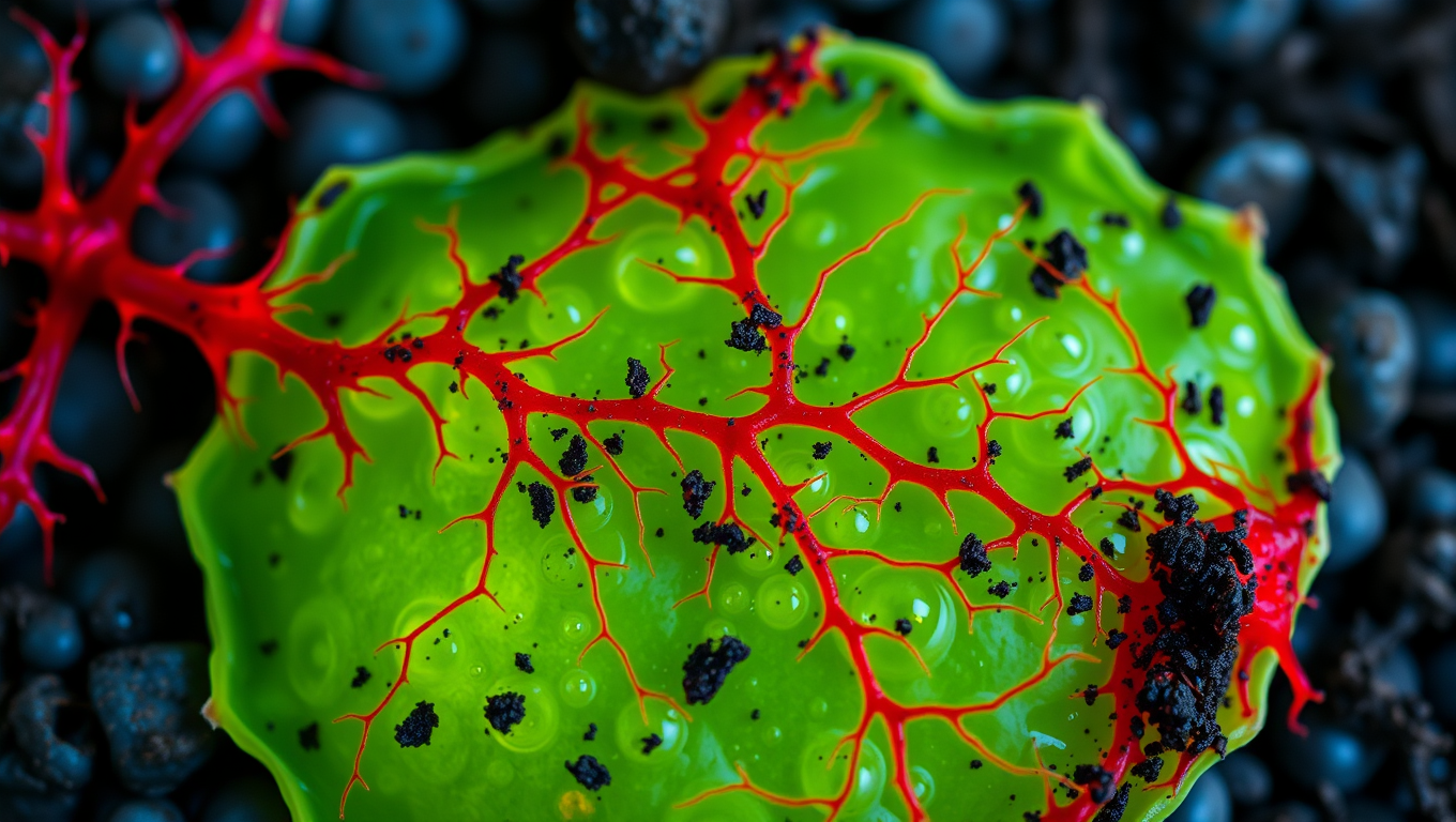
The phenomenon of young organisms getting sick more easily than adults has long puzzled parents and scientists alike. A new study published in the Proceedings of the National Academy of Sciences reveals that young plants may be more vulnerable to disease due to a hidden trade-off involved in fighting off pathogens at an early age. University of Maryland biologists have discovered that baby plants with stronger disease resistance pay a higher “cost” for fighting off diseases, which prevents them from being able to completely fight off the infection.

The researchers studied a wild plant called Silene latifolia and its relationship with a fungal disease called anther-smut. This disease doesn’t kill the plants but prevents them from producing pollen, making them unable to reproduce. By testing 45 different genetic variations of the Silene plant under controlled settings, the team found that plants with stronger disease resistance as seedlings produced significantly fewer flowers and seeds over their lifetime when grown in a disease-free field.

The study suggests that trying to fight off the fungus is more difficult and resource-consuming for young baby plants. They only have so much energy to spend, and if they use it on disease defense, they can’t put it toward future growth. This cost of fighting off pathogens is high enough to prevent the evolution of stronger disease resistance in younger plants.

The researchers created a mathematical model showing that these costs of fighting off pathogens are high enough to prevent the evolution of stronger disease resistance in younger plants. Without these costs, plant families with stronger juvenile resistance would theoretically be able to eliminate the disease entirely. However, because developing resistance is so impactful for young plants, they remain vulnerable to infection.

The team was surprised that these costs didn’t show up right away. Plants that invested in disease resistance as seedlings looked fine at first but produced dramatically fewer flowers in their second year when reproduction would normally peak. Interestingly, the researchers also found that male plants suffered much higher costs for disease resistance than female plants.

Bruns believes that the team’s findings have implications beyond wild plants. Because juvenile susceptibility drives disease epidemics across many species, understanding the evolutionary mechanisms behind this pattern could inform disease management strategies in agriculture, conservation, and public health. The next steps for Bruns and her team include investigating whether disease resistance costs can be reduced by introducing pathogens to plants slightly later in life and exploring whether adult plants with higher disease resistance might protect nearby seedlings.

In conclusion, the hidden cost of young plant disease resistance is a complex phenomenon that requires further investigation. By understanding the evolutionary mechanisms behind this pattern, we may be able to develop more effective strategies for managing diseases across various species.

Biochemistry Research

Unveiling the Origins of HIV Resistance: A 7,000-Year-Old Genetic Mutation Reveals Ancient Secrets

Modern HIV medicine is based on a common genetic mutation. Now, researchers have traced where and when the mutation arose — and how it protected our ancestors from ancient diseases.

Avatar photo

Published

on


The story begins with a simple yet fascinating connection between modern HIV medicine and an ancient human from the Black Sea region. Researchers at the University of Copenhagen have now unraveled the mystery behind a millennia-old genetic mutation that protects against HIV, affecting 18-25% of the Danish population. This breakthrough has shed light on the origins of this crucial genetic variation.

The researchers employed advanced DNA technology to analyze the genetic material of over 2,000 living people worldwide and developed an AI-based method to identify the mutation in ancient DNA from old bones. By examining data from over 900 skeletons dating from the early Stone Age to the Viking Age, they pinpointed the region where the mutation originated – a person from the Black Sea region up to 9,000 years ago.

But why did this genetic mutation arise and spread rapidly among our ancestors? The researchers believe it provided an advantage in surviving during a time when humans were exposed to new pathogens. This variation disrupted an immune gene, which may have been beneficial by dampening the immune system. As humans transitioned from hunter-gatherers to living closely together in agricultural societies, the pressure from infectious diseases increased, and a more balanced immune system may have been advantageous.

The discovery of this 7,000-year-old genetic mutation not only reveals ancient secrets but also provides valuable insights into modern HIV medicine. It highlights the importance of understanding our evolutionary history and how it has shaped our genetic makeup. This breakthrough opens up new avenues for research, potentially leading to innovative treatments for various diseases.

Continue Reading

Biochemistry Research

A More Realistic Look at DNA in Action: Researchers Shed Light on the Mechanical Forces Behind Strand Separation

By creating a more true-to-life representation of DNA’s environment, researchers have discovered that strand separation may take more mechanical force than the field previously believed.

Avatar photo

Published

on

The way we study DNA has long been a topic of discussion among researchers. Traditionally, biochemistry labs isolate DNA within a water-based solution that allows scientists to manipulate it without interacting with other molecules. However, this approach can be misleading, as it doesn’t reflect the true environment of a living cell. In fact, the interior of a cell is “super crowded” with molecules, which can significantly impact the behavior of DNA.

Researchers at Northwestern University have taken a more realistic approach to studying DNA by creating an environment that mimics the conditions within a living cell. Led by Professor John Marko, the team used microscopic magnetic tweezers to separate DNA and then carefully attach strands of it to surfaces on one end, and tiny magnetic particles on the other. This allowed them to conduct high-tech imaging and investigate how different types of molecules interact with DNA.

The researchers found that strand separation, a crucial process for initiating replication or making repairs, may require more mechanical force than previously believed. They introduced three types of molecules to the solution holding DNA, mimicking proteins and investigating interactions among glycerol, ethylene glycol, and polyethylene glycol (each approximately the size of one DNA double helix, two or three nanometers).

“We wanted to have a wide variety of molecules where some cause dehydration, destabilizing DNA mechanically, and then others that stabilize DNA,” said Northwestern post-doctoral researcher Parth Desai. “It’s not exactly analogous to things found in cells, but you could imagine that other competing proteins in cells will have a similar effect.”
The team wrote a paper on their findings, which will be published on June 17 in the Biophysical Journal. Marko and Desai hope to run more experiments that incorporate multiple crowding agents and move closer to a true representation of a cell.

“If this affects DNA strand separation, all protein interactions with DNA are also going to be affected,” said Marko. “For example, the tendency for proteins to stick to specific sites on DNA and to control specific processes — this is also going to be altered by crowding.”

Their research has significant implications for understanding fundamental biochemical processes and may lead to new medical advances. The team hopes to study how interactions between enzymes and DNA are impacted by crowding in a living cell, which could have far-reaching consequences for our understanding of cellular biology.

This work was supported by the National Institutes of Health (grant R01-GM105847) and by subcontract to the University of Massachusetts Center for 3D Structure and Physics of the Genome (under NIH grant UM1-HG011536).

Continue Reading

Bacteria

Unlocking Efficiency: Researchers Reveal Secrets of Cell Division with Min Proteins

The Min protein system prevents abnormal cell division in bacteria, but is poorly understood. Researchers have uncovered how engineered e.coli bacteria control protein levels for maximum efficiency.

Avatar photo

Published

on

The Min protein system is a complex process that helps bacteria divide evenly and correctly. For decades, scientists have studied this system, but understanding how it works efficiently has been a challenge. Recently, researchers at the University of California San Diego (UCSD) made a groundbreaking discovery that sheds new light on the efficiency of cell division.

The UCSD team developed a way to control Min protein expression levels independently in E. coli cells. This allowed them to observe how different concentrations of Min proteins affect the oscillations between the poles of the cell. The results were surprising: despite varying concentrations, the oscillations remained stable across a wide range, with E. coli producing just the right amount of Min proteins.

This breakthrough is significant because it shows that the Min protein system can efficiently guide division to the correct location without relying on precise control over protein levels. This finding has far-reaching implications for our understanding of cellular organization and function.

The study was published in Nature Physics, a leading scientific journal, and was funded by the National Institutes of Health (NIH). The research team consisted of experts from both physics and chemistry/biochemistry departments at UCSD, highlighting the importance of interdisciplinary collaboration in advancing our knowledge of cellular biology.

Continue Reading

Trending