
While we try to keep things accurate, this content is part of an ongoing experiment and may not always be reliable.
Please double-check important details — we’re not responsible for how the information is used.
Biology
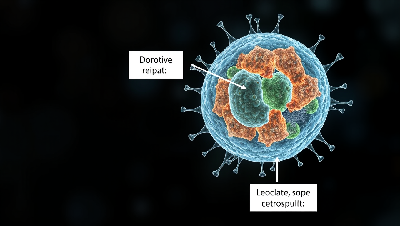
“Uncovering the Cellular Recycling System That Repairs Mitochondrial Power Plants”
Damage to the mitochondria, the ‘power plants’ of the cells, contributes to many diseases. Researchers now describe how cells with defective mitochondria activate a special recycling system to eliminate damaged genetic material.


Behavioral Science
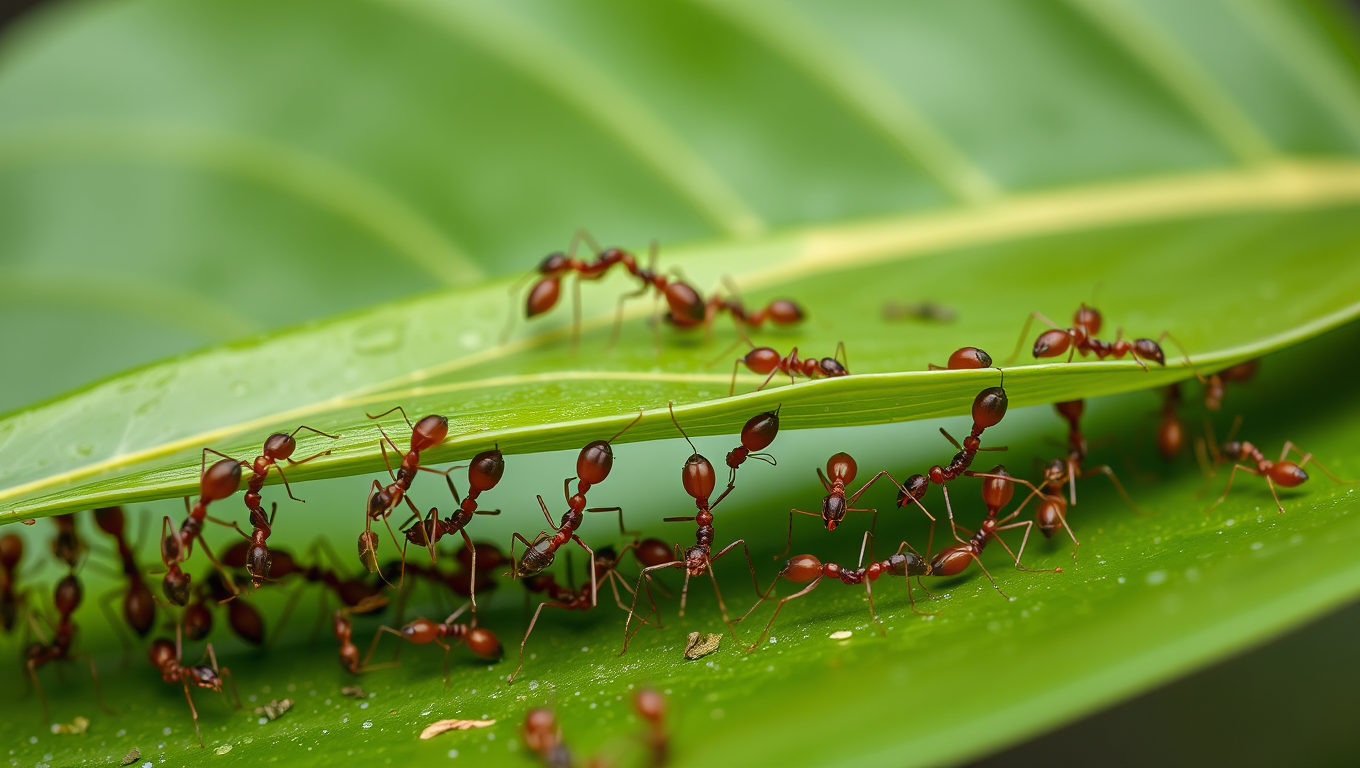
The Amazing Ant Strategy That Can Revolutionize Robotics
Weaver ants have cracked a teamwork puzzle that humans have struggled with for over a century — instead of slacking off as their group grows, they work harder. These tiny architects not only build elaborate leaf nests but also double their pulling power when more ants join in. Using a “force ratchet” system where some pull while others anchor, they outperform the efficiency of human teams and could inspire revolutionary advances in robotics cooperation.
Bacteria
Unlocking the Secrets of Mars: Cosmic Rays Reveal Hidden Potential for Life
Cosmic rays from deep space might be the secret energy source that allows life to exist underground on Mars and icy moons like Enceladus and Europa. New research reveals that when these rays interact with water or ice below the surface, they release energy-carrying electrons that could feed microscopic life, a process known as radiolysis. This breakthrough suggests that life doesn’t need sunlight or heat, just some buried water and radiation.

Animals
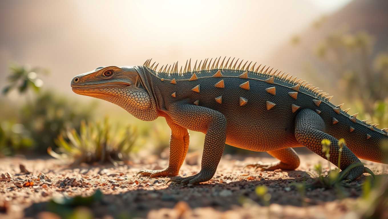
The Hidden Armor of Australia’s Iconic Lizards: Uncovering the Secret Bone Structures that Helped Them Thrive
Scientists have uncovered hidden bony armor—called osteoderms—beneath the skin of 29 goanna species across Australasia, a discovery that radically changes what we thought we knew about lizard evolution. Using museum specimens and advanced scanning, researchers found these structures are far more widespread than previously known, suggesting they may help with survival in harsh environments, not just offer protection. The revelation redefines how we understand lizard adaptation, ancient evolution, and the untapped potential of museum collections.

A New Horizon for Vision: How Gold Nanoparticles May Restore People’s Sight

Revolutionizing Quantum Communication: Direct Connections Between Multiple Processors

Retiring Abroad Can Be Lonely Business
Harnessing Water Waves: A Breakthrough in Controlling Floating Objects

Household Electricity Three Times More Expensive Than Upcoming ‘Eco-Friendly’ Aviation E-Fuels, Study Reveals

“Unveiling Hidden Patterns: A New Twist on Interference Phenomena”
Reducing Falls Among Elderly Women with Polypharmacy through Exercise Intervention
-
 Detectors1 year ago
A New Horizon for Vision: How Gold Nanoparticles May Restore People’s Sight
-
Cancer1 year ago
Revolutionizing Quantum Communication: Direct Connections Between Multiple Processors
-
Earth & Climate1 year ago
Retiring Abroad Can Be Lonely Business
-
Albert Einstein1 year ago
Harnessing Water Waves: A Breakthrough in Controlling Floating Objects
-
Earth & Climate1 year ago
Household Electricity Three Times More Expensive Than Upcoming ‘Eco-Friendly’ Aviation E-Fuels, Study Reveals
-
Chemistry1 year ago
“Unveiling Hidden Patterns: A New Twist on Interference Phenomena”
-
Diseases and Conditions1 year ago
Reducing Falls Among Elderly Women with Polypharmacy through Exercise Intervention
-
Agriculture and Food1 year ago
“A Sustainable Solution: Researchers Create Hybrid Cheese with 25% Pea Protein”


