Connect with us

Animal Learning and Intelligence

Unlocking Brain Health: Boosting Waste Removal System Improves Memory in Old Mice

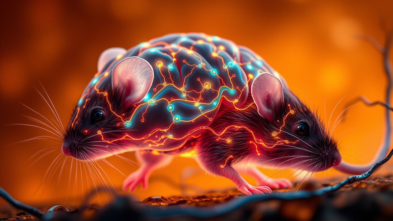
Aging compromises the lymphatic vessels surrounding the brain, disabling waste drainage from the brain and impacting cognitive function. Researchers boosted lymphatic vessel integrity in old mice and found improvements in their memory compared with old mice without rejuvenated lymphatic vessels.
Avatar photo

Published

on

The study, published online March 21 in the journal Cell, reveals that scientists have discovered a way to boost the brain’s waste removal system by targeting the network of vessels that drain waste from the brain. This breakthrough has shown promise in improving memory in old mice.

As aging bodies decline, the brain struggles to cleanse itself of waste, contributing to neurodegenerative conditions such as Alzheimer’s disease and Parkinson’s disease. Researchers at Washington University School of Medicine in St. Louis have found a way around this problem by rejuvenating the vessels that drain waste from the brain.

The study’s first author, Kyungdeok Kim, PhD, a postdoctoral fellow in the Kipnis lab, boosted the functioning of the lymphatic vessels in old mice with a treatment that stimulates vessel growth. He found that older mice with rejuvenated lymphatic vessels spent more time exploring new objects, an indicator of improved memory.

“A functioning lymphatic system is critical for brain health and memory,” said Kim. “Therapies that support the health of the body’s waste management system may have health benefits for a naturally aging brain.”

The researchers also discovered that when the lymphatic system is impaired, the burden of cleaning falls to the brain’s resident immune cells, called microglia. However, this local cleaning crew fails to keep up with the mess and gets exhausted.

The study found that overwhelmed cells produce an immune protein called interleukin 6 (IL-6), which acts on brain cells to promote cognitive decline in mice with damaged lymphatic vessels. Examining the brains of such mice, the researchers found that neurons had an imbalance in the types of signals they receive from surrounding brain cells.

In addition to improving memory in aged mice, the lymphatic vessel-boosting treatment also caused levels of IL-6 to drop, restoring the noise-canceling system of the brain. The findings point to the potential of improving the health of the brain’s lymphatic vessels to preserve or restore cognitive abilities.

“As we mark the 10th anniversary of our discovery of the brain’s lymphatic system, these new findings provide insight into the importance of this system for brain health,” said Jonathan Kipnis, PhD. “Targeting the more easily accessible lymphatic vessels that are located outside the brain may prove to be an exciting new frontier in the treatment of brain disorders.”

The study lays the groundwork for developing therapies that target the brain’s waste removal system, offering hope for future treatments and improved cognitive abilities.

Animal Learning and Intelligence

“Unlocking the Secrets of Animal Energy Consumption: A New Video-Based Method”

Strong methods do exist for measuring animal movement in the context of energy expenditure, but these are limited by the physical size of the equipment used. Now, in a paper published in the Journal of Experimental Biology, researchers from the Marine Biophysics Unit at the Okinawa Institute of Science and Technology (OIST), in collaboration with Professor Amatzia Genin from the Hebrew University of Jerusalem, describe an innovative method for measuring energy usage during movement with video and 3D-tracking via deep learning.

Avatar photo

Published

on

The amazing diversity of life on our planet is a testament to the multitude of biological solutions that have evolved to secure and maintain energy. However, despite its central role in biology, measuring energy consumption remains a challenging task. One major drain for many animals is movement, making it an ideal lens through which to estimate energy usage. While methods exist for measuring animal movement, they are often limited by the physical size of the equipment used.

In a groundbreaking study published in the Journal of Experimental Biology, researchers from the Marine Biophysics Unit at the Okinawa Institute of Science and Technology (OIST) have developed an innovative method for measuring energy usage during movement using video and 3D-tracking via deep learning. This new approach opens up the possibility of studying energy consumption in animals that were previously inaccessible due to the reliance on wearable equipment.

The current state-of-the-art method, Dynamic Body Acceleration (DBA), involves measuring oxygen consumption while an animal performs a specific behavior in a lab setting. However, this method has limitations when applied in the wild, where reliably measuring oxygen consumption is impossible. To overcome these challenges, researchers have used physical accelerometers that weigh at least ten times less than the animal, but this still rules out the study of many small species.

The OIST researchers’ solution to this problem is elegantly simple: they use two cameras to capture video footage of an animal’s behavior from multiple angles, reconstructing its movement in 3D space. A deep learning neural network is then trained on a few frames of the videos to track the position of body features such as eyes, allowing researchers to subsequently measure the movement-related acceleration.

This new video-based DBA method has opened up possibilities for studying energy consumption in animals that were previously inaccessible, potentially enabling many new research avenues into the breadth of life on our planet. For example, researchers can now investigate the energy expenditure during schooling of small fish, which has long remained mysterious. By accurately measuring energy usage during free-ranging animal behavior, scientists can gain a deeper understanding of the ecology and evolution of various species.

Continue Reading

Animal Learning and Intelligence

Mapping the Mouse Brain: Unveiling the Secrets of Visual Perception and Connections

In a massive scientific effort, hundreds of researchers have helped to map the connections between hundreds of thousands of neurons in the mouse brain and then overlayed their firing patterns in response to visual stimuli. This breakthrough is a critical piece of foundational science to build toward understanding how our brains process visual information to reconstruct the images we see every day.

Avatar photo

Published

on

The scientists have made a groundbreaking discovery in understanding the intricacies of the mouse brain. In an unprecedented effort, hundreds of researchers worked together to map the connections between hundreds of thousands of neurons in the mouse brain and overlay their firing patterns in response to visual stimuli. This breakthrough is a crucial piece of foundational science that will help us comprehend how our brains process visual information, allowing us to reconstruct the images we see every day.

The human brain contains 86 billion neurons that make trillions of connections with each other through electrical firings. The complexity of its wiring diagram and the rapid movement of electrical signals across it in millisecond time frames hold the secrets of how our brain enables us to think, feel, and act. Although the current findings focus on a tiny fraction of the brain, they reveal the complex connections between cells and show how those connections are wired to produce functional responses.

To achieve this study, researchers presented video clips to mice genetically engineered for their neurons to emit light when they fire. The neuron firing patterns in areas associated with vision were optically recorded across a cubic millimeter – about the size of a grain of sand. Within this deceptively small amount of tissue lies remarkable complexity: four kilometers of axons, the processes that nerve cells use to communicate with each other, intertwined to make more than 524 million connections called synapses across more than 200,000 cells.

To map these connections, teams worked 12-hour shifts for 12 straight days to carefully cut and image ultra-thin slices of the brain tissue using electron microscopes. Reconstruction was the most challenging next step, as it required accurate stitching together almost 28,000 EM images to align the connections that cross the volume of brain tissue. This was followed by months of tracing the connections using deep learning algorithms, manual proofreading, and automated validation.

Deep learning predictive models were constructed and validated to explain visual information processing in the cortex. The sheer amount of data collected to create this tiny map comes out to 1.6 petabytes, roughly the equivalent of 22 years of continuous HD video.

These results come at a time when maps of neurons and their connections are increasingly revealing the mysteries of the brain. In 2023, research funded by the National Institutes of Health Brain Research Through Advancing Innovative Neurotechnologies Initiative produced the first complete cell atlas of the mouse brain, including the types and locations surveyed from more than 32 million cells. Last year, the NIH BRAIN Initiative “Flywire” project led to the complete mapping of the common fruit fly brain, demonstrating the unique value of mapping the whole brain in its entirety.

Funding for this project was provided through the Machine Intelligence from Cortical Networks Program of the Intelligence Advanced Research Projects Activity and the NIH BRAIN Initiative. The findings, published in a package of 10 papers published in the Nature family of journals, represent more than seven years of work performed by more than 150 scientists around the world.

Continue Reading

Animal Learning and Intelligence

The Link Between Cannabis Use and Psychosis: Uncovering the Brain’s Dopamine System

A new study found that people with cannabis use disorder (CUD) had elevated dopamine levels in a brain region associated with psychosis.

Avatar photo

Published

on

The connection between cannabis use and psychosis has long been a topic of interest for researchers. A recent study led by McGill University sheds light on the brain’s dopamine system as a possible explanation for why cannabis use increases the risk of hallucinations and delusions, key symptoms of schizophrenia and other psychotic disorders.

Dopamine is a neurotransmitter that regulates mood and motivation, and an excess is associated with psychosis. The study reveals that individuals with cannabis use disorder (CUD) have elevated dopamine levels in a brain region linked to psychosis. This finding could help explain why cannabis use increases the risk of hallucinations and delusions.

The study involved 61 participants, including those with and without CUD, as well as individuals with early-stage schizophrenia, some of whom also had CUD. Using a specialized brain scan called neuromelanin-MRI, researchers measured their neuromelanin signal, which reflects dopamine activity. The results showed that people with CUD had an abnormally high neuromelanin signal, and the elevation was tied to the severity of their cannabis use.

These findings have significant implications for educating youth about the risks associated with frequent cannabis use. In Canada, about one-in-five youth are cannabis users, consuming it daily or almost daily. Understanding the potential impact on mental health remains a pressing question.

The study’s lead author, Jessica Ahrens, notes that “for a long time, clinical researchers across the world have been searching for a link showing that cannabis affects the brain mechanism behind psychosis. We now show that a shared dopamine pathway could be the answer.”

Future research will explore whether long-term cannabis use leads to lasting dopamine changes and whether these effects reverse after quitting. As Dr. Lena Palaniyappan, Professor of Psychiatry at McGill and Psychiatrist at the Douglas Mental Health University Institute, suggests, “our findings could help doctors and mental health professionals better educate patients about the potential risks of frequent cannabis use, especially for those with a family history of psychosis.”

The study’s publication in Jama Psychiatry provides valuable insights into the link between cannabis use and psychosis, and its findings have important implications for public health and education.

Continue Reading

Trending