Connect with us
We’re experimenting with AI-generated content to help deliver information faster and more efficiently.
While we try to keep things accurate, this content is part of an ongoing experiment and may not always be reliable.
Please double-check important details — we’re not responsible for how the information is used.

Cell Biology

Unlocking the Secrets of Deadly Tropical Diseases: A New Target for Treatment

The efforts of a research team give hope for new treatment approaches for dangerous tropical diseases. The researchers have compiled a high-precision inventory of the membrane proteins of cell organelles of the African sleeping sickness pathogen.
Avatar photo

Published

on

The fight against deadly tropical diseases has taken a significant step forward with the discovery of a promising target for new treatments. Researchers from Bochum and Würzburg have made a groundbreaking find in their study on the African sleeping sickness pathogen Trypanosoma brucei, which also causes Chagas disease and leishmaniasis.

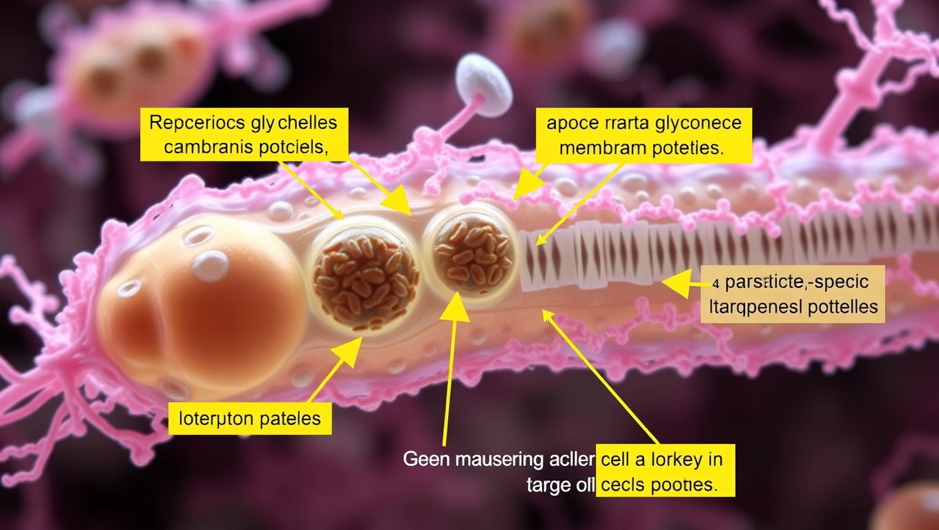
In a breakthrough study published in Cell Reports, the researchers compiled a high-precision inventory of the membrane proteins of the glycosomes, unique cell organelles essential for the survival of the parasites. “Some of these proteins contain components that are specific to parasites and differ significantly from those of the host cells,” explains Professor Ralf Erdmann.

The team’s success in identifying 28 glycosome membrane proteins with a high degree of reliability opens up new avenues for targeted treatment strategies against these poorly understood tropical diseases. A particular highlight was the discovery of TbPEX15, a membrane anchor for an essential protein import complex that differs significantly from its counterpart in humans.

This finding provides a valuable resource for biomedical research into glycosome biology and deepens our understanding of parasite biology. The researchers’ work gives hope for new treatment approaches for diseases that affect over 12 million people worldwide.

The study’s findings are a significant step forward in the fight against deadly tropical diseases, and further research could lead to the development of new therapies and treatments.

Behavioral Science

The Sugar that Sparked Life: Unraveling the Mystery of Ribose’s Preeminence in RNA Development

What made ribose the sugar of choice for life’s code? Scientists at Scripps Research may have cracked a major part of this mystery. Their experiments show that ribose binds more readily and selectively to phosphate compared to other similar sugars, forming a structure ideal for RNA formation. This discovery hints at how nature might have selected specific molecules long before enzymes or life existed, and could reshape our understanding of life’s chemical origins.

Avatar photo

Published

on

The study published in Angewandte Chemie sheds light on how ribose may have become the preferred sugar for RNA development, highlighting its unique ability to bind with phosphate more quickly and effectively than other sugar molecules. This characteristic could have played a crucial role in selecting ribose as the building block of life.

Ramanarayanan Krishnamurthy, professor of chemistry at Scripps Research, emphasizes that this finding supports the idea that prebiotic chemistry could have produced the fundamental components of RNA, which eventually led to entities exhibiting lifelike properties. The research focuses on phosphorylation, a step within nucleotide-building where ribose connects to the phosphate group, and explores whether other sugars can undergo similar reactions.

The team’s experiments showed that while diamidophosphate (DAP) could phosphorylate all four sugar molecules tested, it phosphorylated ribose at a significantly faster rate. The reaction with ribose produced exclusively ring-shaped structures with five corners, whereas the other sugars formed a combination of 5- and 6-member rings.

“This really showed us that there is a difference between ribose and the three other sugars,” says Krishnamurthy. “Ribose not only reacts faster than the other sugars, it’s also more selective for the five-member ring form, which happens to be the form that we see in RNA and DNA today.”

When DAP was added to a solution containing equal amounts of the four different sugars, it preferentially phosphorylated ribose. The researchers demonstrated that this selective process produces a molecule with a form conducive for making RNA, providing further evidence for ribose’s preeminence.

While the study does not claim that these reactions directly led to life, it suggests that they might have played a crucial role in the primordial process that gave rise to the fundamental components of life. The researchers caution against over-interpretation and emphasize the need for further investigation into the emergence of life on Earth.

In future research, the team plans to test whether this chemical reaction can occur inside primitive cellular structures called protocells. If successful, it might provide a compelling explanation for how ribose became the preferred sugar for RNA development and ultimately gave rise to life as we know it today.

Continue Reading

Cell Biology

A 600-Million-Year-Old Body Blueprint Uncovered in Sea Anemones

Sea anemones may hold the key to the ancient origins of body symmetry. A study from the University of Vienna shows they use a molecular mechanism known as BMP shuttling, once thought unique to bilaterally symmetrical animals like humans, insects, and worms. This surprising discovery implies that the blueprint for forming a back-to-belly body axis could date back over 600 million years, to a common ancestor of cnidarians and bilaterians.

Avatar photo

Published

on

A new study from the University of Vienna has made a groundbreaking discovery in the field of developmental biology. Researchers have found that sea anemones, traditionally considered radially symmetric animals, use a molecular mechanism known as BMP shuttling to pattern their back-to-belly body axis. This finding suggests that bilateral symmetry, which characterizes a vast group of animals including vertebrates, insects, and worms, may have evolved much earlier than previously assumed.

BMP shuttling is a signaling system involving Bone Morphogenetic Proteins (BMPs) and their inhibitor Chordin. In bilaterian animals, this mechanism creates a gradient of BMP activity across the embryo, allowing cells to detect and adopt different fates depending on BMP levels. The study’s findings indicate that sea anemones use BMP shuttling in a similar manner, with cells expressing different fates based on BMP signaling.

To investigate whether sea anemones indeed use BMP shuttling, researchers blocked Chordin production in the embryos of the model sea anemone Nematostella vectensis. Without Chordin, BMP signaling ceased, and the formation of the second body axis failed. However, when Chordin was reintroduced into a small part of the embryo, BMP signaling resumed – but only with a diffusible form of Chordin, which acts as a BMP shuttle.

The presence of BMP shuttling in both cnidarians and bilaterians suggests that this molecular mechanism predates their evolutionary divergence some 600-700 million years ago. The study’s findings open up exciting possibilities for rethinking how body plans evolved in early animals, and may have significant implications for our understanding of the evolution of bilateral symmetry.

The research was supported by the Austrian Science Fund (FWF), grants P32705 and M3291.

Continue Reading

Biology

Unraveling Microtubule Mysteries: Scientists Crack Code on Cellular Scaffolding Secrets

Scientists found out how naturally unstable filaments decide whether to grow or to shorten.

Avatar photo

Published

on

A groundbreaking study has shed new light on the fundamental mechanisms governing microtubule growth within cells. Researchers from Queen Mary University of London and the University of Dundee have made a significant breakthrough by discovering that the ability of tubulin proteins at microtubule ends to connect with each other sideways determines whether a microtubule elongates or shortens.

Microtubules are crucial protein structures that form the internal skeleton of cells, providing structural support and generating dynamic forces that push and pull. These tiny filaments constantly assemble and disassemble by adding or removing tubulin building blocks at their ends. However, the precise rules dictating whether a microtubule grows or shrinks have long remained a mystery due to the complexity and miniature size of their ends.

The collaborative research team has cracked part of this code using advanced computer simulations coupled with innovative imaging techniques. This interdisciplinary approach has allowed them to address this complex biological question from a fresh perspective, bridging physics and biology.

Dr. Vladimir Volkov, co-lead author from Queen Mary University of London, explained the significance of their findings: “Understanding how microtubules grow and shorten is very important – this mechanism underlies division and motility of all our cells. Our results will inform future biomedical research, particularly in areas related to cell growth and cancer.”

Dr. Maxim Igaev, co-lead author from the University of Dundee, highlighted the power of their interdisciplinary approach: “Bridging physics and biology has allowed us to address this complex biological question from a fresh perspective. This synergy not only enriches both fields but also paves the way for discoveries that neither discipline could achieve in isolation.”

This exciting research deepens our understanding of fundamental cellular processes and opens potential new avenues for biomedical research, particularly in areas concerning cell proliferation and the development of treatments for diseases like cancer.

Continue Reading

Trending