Connect with us

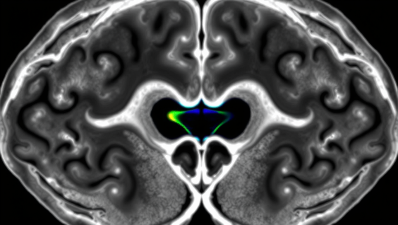
Brain-Computer Interfaces

Unlocking the Secrets of the Brainstem: A Breakthrough in Understanding Brain-Body-Mind Interactions

Researchers have developed a new imaging method, D-PSCAN, which enables minimally invasive, wide-field, high-resolution imaging of the nucleus tractus solitarii (NTS) in living mice. This technique allows detailed investigation of NTS activity and offers broad potential for advancing our understanding of brain–body–mind interactions, as well as informing therapeutic strategies for psychiatric and neurological disorders.

Avatar photo

Published

on

The brain’s communication with bodily organs is a crucial aspect of emotion regulation and overall mental health. The nucleus tractus solitarii (NTS), located in the brainstem, plays a vital role as a hub structure mediating this interaction through the vagus nerve. Despite its importance, the NTS’s deep location has historically presented challenges for observation in living animals.

A recent study published in Cell Reports Methods (April 4, 2025) has made significant strides in overcoming these challenges by developing a novel live imaging technique called D-PSCAN. This minimally invasive method enables high-resolution visualization of the NTS neural activity in living mice, offering unprecedented insights into its function.

The D-PSCAN technique involves implanting a double microprism assembly between the cerebellum and brainstem, preserving cerebellar function while providing a detailed view of the NTS. This approach overcomes the major limitation of previous methods, which often involved removing the cerebellum to access the NTS, thus compromising its role in emotional regulation.

The research team evaluated the D-PSCAN method by investigating the NTS’s response to electrical stimulation of the vagus nerve, which conveys signals from internal organs to the NTS. They observed specific thresholds of vagus nerve stimulation intensity required to elicit neural responses in the NTS and noted distinct patterns of neural activation upon varying stimulation parameters.

These results have significant implications for therapeutic applications, particularly for vagus nerve stimulation (VNS), which has been used clinically for drug-resistant epilepsy and is currently under investigation as a treatment for depression and other psychiatric and neurological disorders. The D-PSCAN method offers valuable insights into optimizing VNS parameters for maximum effectiveness.

To further explore NTS function under more physiological conditions than electrical stimulation, the research team applied the D-PSCAN method to examine its response to the gut hormone cholecystokinin, which is naturally released after feeding. They successfully detected NTS neural activity evoked by cholecystokinin, providing new insights into the complex interactions between the brain and body.

The implications of this research extend beyond the study of emotion regulation, as the NTS receives input from various organs, including the heart and gut, and is involved in diverse functions such as appetite regulation, energy metabolism, and gut microbiota. The D-PSCAN technique is expected to be widely applied across these research areas, offering a new approach to elucidate brain-body-mind interactions and contributing both to the treatment of neuropsychiatric disorders and to the advancement of mental health and well-being.

Behavior

The Adaptive Nature of Object Recognition: How Experience Shapes Our Perception of Reality

New findings suggest neurons have much more functional dexterity than scientists previously realized.

Avatar photo

Published

on

By

The human brain has an incredible ability to recognize objects from a very young age. This process involves the visual cortex, which is responsible for processing visual information from the eyes. While it’s been thought that specific neurons along this pathway handle specific types of information depending on their location, new research suggests that feedback connections play a crucial role in object recognition. These connections convey information from higher cortical areas to lower ones, contributing to the dynamic capabilities of the brain.

Studies have shown that even at the first stages of object perception, neurons are sensitive to much more complex visual stimuli than previously believed. This capability is informed by feedback from higher cortical areas, which can adapt moment-to-moment to the information they’re receiving. In fact, researchers have found that a single neuron may be more responsive to one target and with another cue, they’ll be more responsive to a different target.

This adaptive processing allows the brain to dynamically tune its functional properties, changing its specificities with varying sensory experience. The findings of this research have significant implications for our understanding of how we perceive reality and could lead to new insights into the mechanisms underlying brain disorders such as autism.

In fact, researchers are now beginning to investigate animal models of autism at both the behavioral and imaging level. By studying perceptual differences between autism-model mice and their wild-type littermates, scientists hope to identify any cortical circuitry differences that may underlie these differences.

Overall, this research highlights the complex and dynamic nature of object recognition processes in the brain, and how experience shapes our perception of reality. It also underscores the importance of feedback connections in this process and has significant implications for our understanding of brain function and disorders.

Continue Reading

Brain Tumor

Hair-like Electrodes Revolutionize Brain Activity Monitoring

Researchers have created a hairlike device for long-term, non-invasive monitoring of the brain’s electrical activity. The lightweight and flexible electrode attaches directly to the scalp and delivers stable, high-quality electroencephalography (EEG) recordings.

Avatar photo

Published

on

The article you provided has been rewritten for clarity, structure, and style, making it accessible to a general audience. Here’s the rewritten content:

In the realm of electroencephalography (EEG) monitoring, researchers at Penn State have made a groundbreaking discovery – one that could revolutionize the way we monitor brain activity. Gone are the days of cumbersome metal electrodes; instead, a team of scientists has created hair-like devices for non-invasive, long-term monitoring.

The innovative electrode is designed to mimic human hair and can be worn without drawing attention. This lightweight and flexible device captures stable, high-quality recordings of the brain’s signals for over 24 hours of continuous wear. The traditional metal electrodes used in EEG monitoring are rigid and can shift when someone moves their head, compromising data uniformity.

The new electrode uses a 3D-printed bioadhesive ink that allows it to stick directly onto the scalp without any gloopy gels or skin preparation. This minimizes the gap between the electrode and skin, improving signal quality. The device is also stretchable, ensuring it stays put even when combing hair or wearing a baseball cap.

The researchers found that the new device performed comparably to gold electrodes, the current standard for EEG monitoring. However, the hair-like electrode maintained better contact between the electrode and skin and performed reliably for extended periods without any degradation in signal quality.

According to Tao Zhou, Wormley Family Early Career Professor of Engineering Science and Mechanics, this technology holds promise for use in consumer health and wellness products, as well as clinical healthcare applications.

The conventional EEG monitoring process can be a cumbersome affair, requiring the application of gels to maintain good surface-to-surface contact between the electrodes and skin. This process is imprecise and can result in different amounts of gel used on the electrodes, affecting brain signal quality.

Zhou explained that this new device will change the impedance – or interface – between the electrodes and scalp, ensuring more consistent and reliable monitoring of EEG signals. The researchers also hope to make the system wireless in the future, allowing people to move around freely during recording sessions.

The team’s findings were published in a study in npc biomedical innovations, with funding from various institutions, including the National Institutes of Health and Oak Ridge Associated Universities.

In conclusion, the development of hair-like electrodes for brain activity monitoring is a significant breakthrough that could revolutionize the field. With its potential for non-invasive, long-term monitoring, this technology has far-reaching implications for healthcare and consumer products alike.

Continue Reading

Brain Injury

Uncovering the Aggressive Nature of Glioblastoma: ZIP4’s Role in Brain Tumor Growth

Researchers detail their discoveries about why the brain tumor glioblastoma is so aggressive. Their findings center on ZIP4, a protein that transports zinc throughout the body and sets off a cascade of events that drive tumor growth.

Avatar photo

Published

on

In a groundbreaking study published in the Proceedings of the National Academy of Sciences (PNAS), University of Oklahoma researchers have made a significant discovery about what makes glioblastoma, the deadliest form of brain cancer, so aggressive. The findings center on ZIP4, a protein that transports zinc throughout the body and sets off a chain reaction that drives tumor growth.

Glioblastomas account for about half of all malignant brain tumors, with a median survival rate of 14 months. Surgery is often challenging, and patients almost always experience a relapse. By better understanding why these brain tumors are so aggressive, researchers hope to open up paths for new treatments.

In normal conditions, ZIP4 plays a positive role, transporting and maintaining the right amount of zinc for good health. However, when brain cancer is present, ZIP4 takes on a different role. In the case of glioblastoma, it triggers a series of events that contribute to the tumor’s aggressive growth.

“Everything starts with the fact that ZIP4 is overexpressed in glioblastoma,” says senior author Min Li, Ph.D., a professor of medicine, surgery, and cell biology at the University of Oklahoma College of Medicine. “That triggers all these downstream events that help the tumor to grow.”

Li’s research team tested a small-molecule inhibitor that targets ZIP4 and TREM1, a protein involved in immune responses. The inhibitor attached to both proteins, stopping their actions and slowing tumor growth. This suggests that ZIP4 and TREM1 may be promising therapeutic targets.

Neurosurgeon Ian Dunn, M.D., executive dean of the OU College of Medicine and co-author of the study, says the findings are an encouraging step toward combating this debilitating cancer. “These results are really exciting in such a debilitating cancer. The hope and promise is to translate these findings to novel treatment approaches to improve the lives of our patients.”

This discovery is significant not only for glioblastoma but also for pancreatic cancer research, as ZIP4 has been a focus of Li’s work on this disease for many years. He found that overexpression of ZIP4 causes pancreatic cancer cells to be more resistant to chemotherapy and prompts tumor cells to transform themselves so they can stealthily travel to the body’s other organs.

The researchers hope that their findings will lead to new treatment approaches for glioblastoma and potentially other types of cancer, improving the lives of patients affected by these devastating diseases.

Continue Reading

Trending