Connect with us

Animal Learning and Intelligence

Unlocking the Secrets of the Human Brain: Uncovering the Evolutionary Enlargement of Our Largest Organ

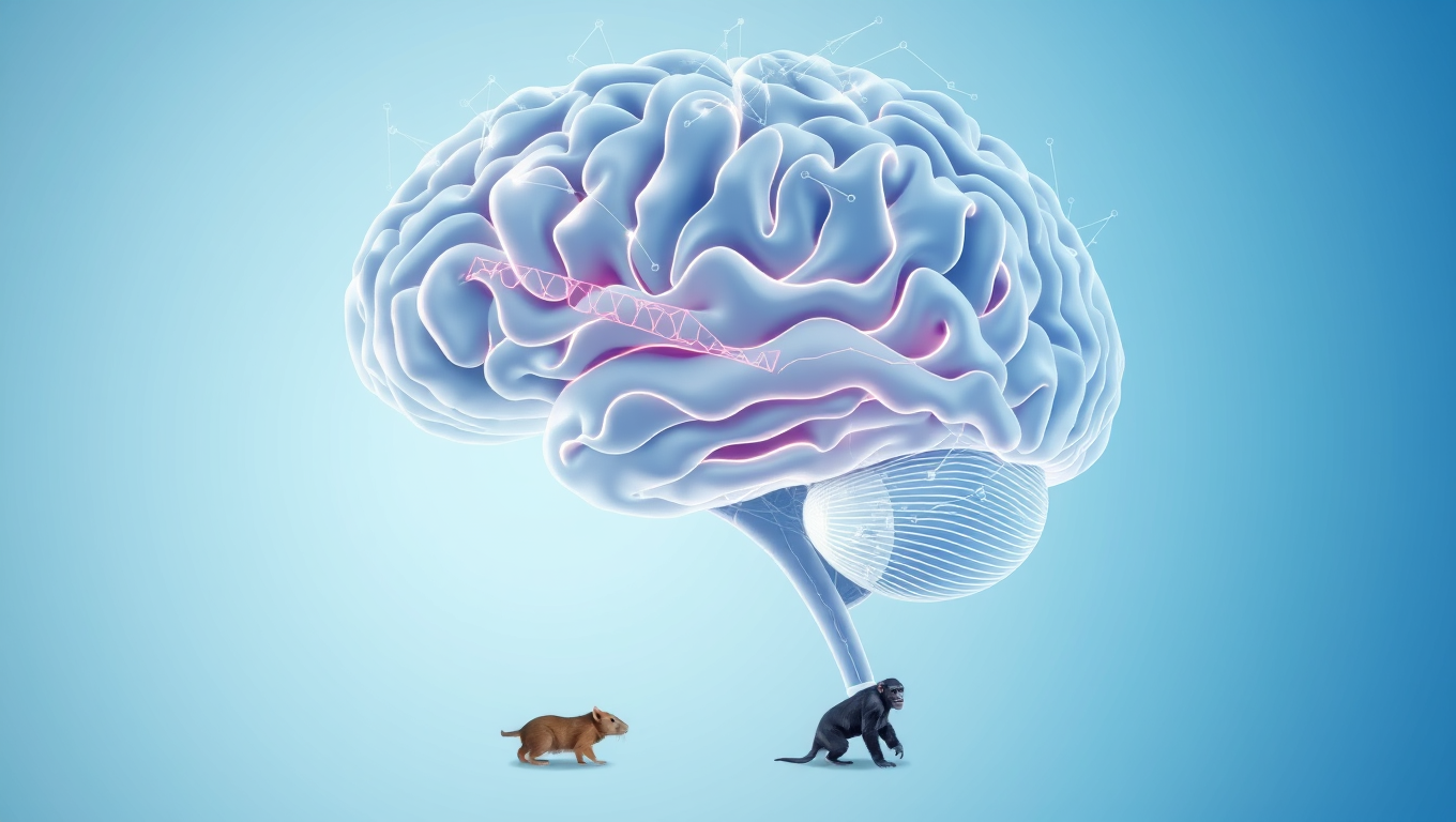
Two specific genes that evolve exclusively in humans jointly influence the development of the cerebrum. Researchers have provided evidence that these genes contribute together to the evolutionary enlargement of the brain.
Avatar photo

Published

on

The human brain is a marvel of complexity, containing over 100 billion neurons that enable us to think, learn, and interact with the world around us. But have you ever wondered how our brains evolved to become so large and sophisticated? A recent study by researchers from the German Primate Center and the Max Planck Institute has shed new light on this question, providing evidence that two specific genes work together to shape the development of our cerebrum.

These genes, which are unique to humans, have a delicate interplay that is crucial for brain growth. One gene ensures that progenitor cells multiply more, while the other causes these cells to transform into nerve cell precursors – the building blocks of the brain. As this process unfolded over millions of years, it led to the human brain becoming distinct in its size and complexity.

The study’s findings are not only fascinating from an evolutionary perspective but also hold promise for better understanding developmental disorders or diseases affecting the brain. According to Nesil Eşiyok, first author of the research, “Our results deepen our fundamental understanding of brain development and provide new insights into the evolutionary origins of our large brain.”

To reach this groundbreaking conclusion, the researchers employed a multi-faceted approach that combined traditional animal experiments with chimpanzee brain organoids – a revolutionary technique that allows scientists to study complex biological systems in a controlled environment. This hybrid method not only validated each other’s findings but also has the potential to reduce the need for animal testing in future research.

The German Primate Center, where this study was conducted, is a world-renowned institution dedicated to advancing our understanding of primates and the human brain. By pushing the boundaries of scientific knowledge, researchers at the center are paving the way for innovative therapeutic approaches that can improve human health and well-being.

Animal Learning and Intelligence

Mapping the Mouse Brain: Unveiling the Secrets of Visual Perception and Connections

In a massive scientific effort, hundreds of researchers have helped to map the connections between hundreds of thousands of neurons in the mouse brain and then overlayed their firing patterns in response to visual stimuli. This breakthrough is a critical piece of foundational science to build toward understanding how our brains process visual information to reconstruct the images we see every day.

Avatar photo

Published

on

The scientists have made a groundbreaking discovery in understanding the intricacies of the mouse brain. In an unprecedented effort, hundreds of researchers worked together to map the connections between hundreds of thousands of neurons in the mouse brain and overlay their firing patterns in response to visual stimuli. This breakthrough is a crucial piece of foundational science that will help us comprehend how our brains process visual information, allowing us to reconstruct the images we see every day.

The human brain contains 86 billion neurons that make trillions of connections with each other through electrical firings. The complexity of its wiring diagram and the rapid movement of electrical signals across it in millisecond time frames hold the secrets of how our brain enables us to think, feel, and act. Although the current findings focus on a tiny fraction of the brain, they reveal the complex connections between cells and show how those connections are wired to produce functional responses.

To achieve this study, researchers presented video clips to mice genetically engineered for their neurons to emit light when they fire. The neuron firing patterns in areas associated with vision were optically recorded across a cubic millimeter – about the size of a grain of sand. Within this deceptively small amount of tissue lies remarkable complexity: four kilometers of axons, the processes that nerve cells use to communicate with each other, intertwined to make more than 524 million connections called synapses across more than 200,000 cells.

To map these connections, teams worked 12-hour shifts for 12 straight days to carefully cut and image ultra-thin slices of the brain tissue using electron microscopes. Reconstruction was the most challenging next step, as it required accurate stitching together almost 28,000 EM images to align the connections that cross the volume of brain tissue. This was followed by months of tracing the connections using deep learning algorithms, manual proofreading, and automated validation.

Deep learning predictive models were constructed and validated to explain visual information processing in the cortex. The sheer amount of data collected to create this tiny map comes out to 1.6 petabytes, roughly the equivalent of 22 years of continuous HD video.

These results come at a time when maps of neurons and their connections are increasingly revealing the mysteries of the brain. In 2023, research funded by the National Institutes of Health Brain Research Through Advancing Innovative Neurotechnologies Initiative produced the first complete cell atlas of the mouse brain, including the types and locations surveyed from more than 32 million cells. Last year, the NIH BRAIN Initiative “Flywire” project led to the complete mapping of the common fruit fly brain, demonstrating the unique value of mapping the whole brain in its entirety.

Funding for this project was provided through the Machine Intelligence from Cortical Networks Program of the Intelligence Advanced Research Projects Activity and the NIH BRAIN Initiative. The findings, published in a package of 10 papers published in the Nature family of journals, represent more than seven years of work performed by more than 150 scientists around the world.

Continue Reading

Animal Learning and Intelligence

The Link Between Cannabis Use and Psychosis: Uncovering the Brain’s Dopamine System

A new study found that people with cannabis use disorder (CUD) had elevated dopamine levels in a brain region associated with psychosis.

Avatar photo

Published

on

The connection between cannabis use and psychosis has long been a topic of interest for researchers. A recent study led by McGill University sheds light on the brain’s dopamine system as a possible explanation for why cannabis use increases the risk of hallucinations and delusions, key symptoms of schizophrenia and other psychotic disorders.

Dopamine is a neurotransmitter that regulates mood and motivation, and an excess is associated with psychosis. The study reveals that individuals with cannabis use disorder (CUD) have elevated dopamine levels in a brain region linked to psychosis. This finding could help explain why cannabis use increases the risk of hallucinations and delusions.

The study involved 61 participants, including those with and without CUD, as well as individuals with early-stage schizophrenia, some of whom also had CUD. Using a specialized brain scan called neuromelanin-MRI, researchers measured their neuromelanin signal, which reflects dopamine activity. The results showed that people with CUD had an abnormally high neuromelanin signal, and the elevation was tied to the severity of their cannabis use.

These findings have significant implications for educating youth about the risks associated with frequent cannabis use. In Canada, about one-in-five youth are cannabis users, consuming it daily or almost daily. Understanding the potential impact on mental health remains a pressing question.

The study’s lead author, Jessica Ahrens, notes that “for a long time, clinical researchers across the world have been searching for a link showing that cannabis affects the brain mechanism behind psychosis. We now show that a shared dopamine pathway could be the answer.”

Future research will explore whether long-term cannabis use leads to lasting dopamine changes and whether these effects reverse after quitting. As Dr. Lena Palaniyappan, Professor of Psychiatry at McGill and Psychiatrist at the Douglas Mental Health University Institute, suggests, “our findings could help doctors and mental health professionals better educate patients about the potential risks of frequent cannabis use, especially for those with a family history of psychosis.”

The study’s publication in Jama Psychiatry provides valuable insights into the link between cannabis use and psychosis, and its findings have important implications for public health and education.

Continue Reading

Animal Learning and Intelligence

“The Optimism Effect: How Medical Detection Dogs’ Emotional States Influence Their Performance”

A new, exploratory study has revealed statistical links between the performance of medical detection dogs and their scores on behavioral and affective tests, finding that more ‘optimistic’ dogs tended to perform better overall on detection tasks, but ‘pessimistic’ dogs had higher scent detection specificity.

Avatar photo

Published

on

The article’s original findings have been rewritten to improve clarity, structure, and style while maintaining its core ideas. Here’s the rewritten version:

Researchers at the University of Bristol recently conducted a groundbreaking study on medical detection dogs, uncovering statistical links between their performance in scent detection tasks and their emotional states. The study, published in PLOS One, found that “optimistic” dogs tended to perform better overall on detection tasks, while “pessimistic” dogs had higher specificity in scent detection.

To assess the dogs’ emotional states, researchers used a behavioral test called judgment bias testing. In this test, dogs are first trained to associate specific locations with treats and others without treats. When presented with intermediate locations where a treat may or may not be present, the dogs’ responses are evaluated as a proxy for their emotional states.

More “optimistic” dogs, characterized by quicker approaches to intermediate locations in hopes of a treat, tended to score higher on detection tasks. These dogs also showed higher confidence, food orientation, and playfulness scores. In contrast, more “pessimistic” dogs achieved a higher degree of specificity in scent detection tasks among the fully trained medical detection dogs.

This study does not establish cause-effect relationships, but it suggests that differences in dogs’ searching styles and performance in detection tasks could be influenced by underlying differences in affective or cognitive processes. The researchers propose that judgment bias testing might hold promise as a screening tool for potential detection dogs.

The findings have significant implications for training and selection methods in medical detection dogs. Understanding the link between emotional states and performance can help trainers select and support successful dogs. The study’s insights could also be relevant to the welfare of companion dogs, highlighting the importance of considering their emotional well-being in their care and management.

Continue Reading

Trending