Connect with us

Brain Injury

Uncovering the Secrets of Thirst and Hunger Neurons: A New Frontier in Brain Research

New research shines light on how the brain interprets nutritional and hydration needs and turns them into action.
Avatar photo

Published

on

Uncovering the Secrets of Thirst and Hunger Neurons: A New Frontier in Brain Research

In order to survive, our bodies require the right balance of hydration and nutrition. To ensure this delicate balance, the brain relies on a complex network of cells, signals, and pathways that guide us when to eat, drink, or stop. Despite its importance, much about how the brain deciphers the body’s needs and translates them into action remains unknown. Recently, researchers from the Max Planck Institute for Biological Intelligence, in collaboration with the University of Regensburg and Stanford University, made a groundbreaking discovery – identifying specific populations of neurons in the amygdala that play a key role in regulating our nutritional needs.

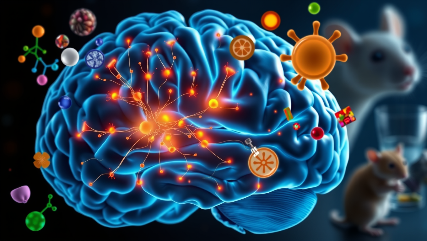
These specialized “thirst” and “hunger” neurons operate through distinct circuits, influencing the drive to eat or drink. The study, conducted in mice, sheds new light on the amygdala’s role in regulating our nutritional needs and may offer insights into eating disorders and addiction.

The amygdala, a brain region often linked to emotions and decision making, also plays a key role in shaping our desire to eat and drink. Earlier research led by Rüdiger Klein’s group at the Max Planck Institute for Biological Intelligence revealed that neurons in the central nucleus of the amygdala connect food to feelings – pairing tasty meals with positive emotions, associating bad food with aversion, and suppressing appetite when nausea sets in.

Building on these findings, the new research has detailed distinct groups of neurons in the same central region of the amygdala that respond specifically to thirst and others that respond to hunger. These neurons are guided by a complex web of molecular cues. “One of these groups of neurons is solely dedicated to regulating the desire to drink, the first ‘thirst neuron’ that has been identified in the amygdala,” explains Federica Fermani, who led the study.

The researchers used advanced genetic tools to study brain activity in mice during hunger, thirst, and when they were already full and hydrated. One method, called optogenetics, allowed them to activate specific neurons using light-sensitive proteins and a laser precisely tuned to trigger those cells. They also used approaches to silence the neurons, observing how their absence influenced the mice’s tendency to eat or drink.

By combining this with new methods that enable the monitoring of individual neurons across multiple brain regions, the researchers mapped where these neurons receive information and identified other brain regions they communicate with. This revealed connections to areas involved in processing sensory information about food and water, such as the parabrachial complex.

The study also explored how the brain balances other factors, such as taste, in shaping behavior. For example, by pairing a less-preferred drink flavor with targeted stimulation of neurons in the central amygdala, the researchers found they could change the mice’s choices, transforming a previously avoided flavor into a new favorite.

This work opens the door to new questions – such as how the brain balances appetite, thirst, and emotions; how we know when we’ve had too little or too much to eat and drink; how competing needs are managed simultaneously; and how these circuits are affected in conditions like obesity, anorexia, or alcohol addiction.

In conclusion, this groundbreaking research has shed new light on the complex neural mechanisms involved in regulating thirst and hunger. As our understanding of these processes grows, we may gain valuable insights into eating disorders and other neurological conditions, ultimately leading to improved treatments and a better quality of life for individuals affected by these conditions.

Biochemistry

A Breakthrough in Brain Research: The Iontronic Pipette Revolutionizes Neurological Studies

Researchers have developed a new type of pipette that can deliver ions to individual neurons without affecting the sensitive extracellular milieu. Controlling the concentration of different ions can provide important insights into how individual brain cells are affected, and how cells work together. The pipette could also be used for treatments.

Avatar photo

Published

on

The development of an iontronic pipette at Linköping University has opened up new avenues for neurological research. This innovative tool allows researchers to deliver ions directly to individual neurons without affecting the surrounding extracellular milieu. By controlling the concentration of various ions, scientists can gain valuable insights into how brain cells respond to different stimuli and interact with each other.

The human brain consists of approximately 85-100 billion neurons, supported by a similar number of glial cells that provide essential functions such as nutrition, oxygenation, and healing. The extracellular milieu, a fluid-filled space between the cells, plays a crucial role in maintaining cell function. Changes in ion concentration within this environment can activate or inhibit neuronal activity, making it essential to study how local changes affect individual brain cells.

Previous attempts to manipulate the extracellular environment involved pumping liquid into the area, disrupting the delicate biochemical balance and making it difficult to determine whether the substances themselves or the changed pressure were responsible for the observed effects. To overcome this challenge, researchers at the Laboratory of Organic Electronics developed an iontronic micropipette measuring only 2 micrometers in diameter.

This tiny pipette can deliver ions such as potassium and sodium directly into the extracellular milieu, allowing scientists to study how individual neurons respond to these changes. Glial cell activity is also monitored, providing a more comprehensive understanding of brain function.

Theresia Arbring Sjöström, an assistant professor at LOE, highlighted that glial cells are critical components of the brain’s chemical environment and can be precisely activated using this technology. In experiments conducted on mouse hippocampus tissue slices, it was observed that neurons responded dynamically to changes in ion concentration only after glial cell activity had saturated.

This research has significant implications for neurological disease treatment. The iontronic pipette could potentially be used to develop extremely precise treatments for conditions such as epilepsy, where brain function can be disrupted by localized imbalances in ion concentrations.

Researchers are now continuing their studies on chemical signaling in healthy and diseased brain tissue using the iontronic pipette. They also aim to adapt this technology to deliver medical drugs directly to affected areas of the brain, paving the way for more targeted treatments for neurological disorders.

Continue Reading

Brain Injury

“Resonating with Sound: Study Suggests Our Brains Physically Respond to Music”

Psychologists suggest our brains and bodies don’t just understand music, they physically resonate with it. These discoveries, based on findings in neuroscience, music, and psychology, support Neural Resonance Theory (NRT).

Avatar photo

Published

on

By

A groundbreaking study co-authored by McGill psychologist Caroline Palmer has revealed that our brains and bodies don’t just understand music; they physically resonate with it. This revolutionary discovery is based on findings in neuroscience, music, and psychology, supporting the Neural Resonance Theory (NRT).

According to NRT, musical experiences arise from the brain’s natural oscillations that sync with rhythm, melody, and harmony. This resonance shapes our sense of timing, musical pleasure, and the instinct to move with the beat.

“This theory suggests that music is powerful not just because we hear it, but because our brains and bodies become it,” said Palmer, Professor in the Department of Psychology at McGill and Director of the Sequence Production Lab. “That has big implications for therapy, education, and technology.”

The study’s publication in Nature Reviews Neuroscience marks the first time the entire NRT is being published in a single paper, she added.

NRT suggests that structures like pulse and harmony reflect stable resonant patterns in the brain, shared across people independent of their musical background. This theory explains how we hear and produce music through fundamental dynamical principles of human brain mechanisms that apply from the ear to the spinal cord and limb movements.

The study’s findings have significant potential applications:

Therapeutic tools for conditions like stroke, Parkinson’s, and depression

Emotionally intelligent AI that can respond to or generate music more like humans

New learning technologies to support rhythm and pitch education

Cross-cultural insight into why music connects people around the world

The study was led by Edward Large (University of Connecticut) and co-authored by Caroline Palmer. The research received funding from a Canada Research Chair and a NSERC Discovery Grant.

Continue Reading

Behavior

The Adaptive Nature of Object Recognition: How Experience Shapes Our Perception of Reality

New findings suggest neurons have much more functional dexterity than scientists previously realized.

Avatar photo

Published

on

By

The human brain has an incredible ability to recognize objects from a very young age. This process involves the visual cortex, which is responsible for processing visual information from the eyes. While it’s been thought that specific neurons along this pathway handle specific types of information depending on their location, new research suggests that feedback connections play a crucial role in object recognition. These connections convey information from higher cortical areas to lower ones, contributing to the dynamic capabilities of the brain.

Studies have shown that even at the first stages of object perception, neurons are sensitive to much more complex visual stimuli than previously believed. This capability is informed by feedback from higher cortical areas, which can adapt moment-to-moment to the information they’re receiving. In fact, researchers have found that a single neuron may be more responsive to one target and with another cue, they’ll be more responsive to a different target.

This adaptive processing allows the brain to dynamically tune its functional properties, changing its specificities with varying sensory experience. The findings of this research have significant implications for our understanding of how we perceive reality and could lead to new insights into the mechanisms underlying brain disorders such as autism.

In fact, researchers are now beginning to investigate animal models of autism at both the behavioral and imaging level. By studying perceptual differences between autism-model mice and their wild-type littermates, scientists hope to identify any cortical circuitry differences that may underlie these differences.

Overall, this research highlights the complex and dynamic nature of object recognition processes in the brain, and how experience shapes our perception of reality. It also underscores the importance of feedback connections in this process and has significant implications for our understanding of brain function and disorders.

Continue Reading

Trending