
While we try to keep things accurate, this content is part of an ongoing experiment and may not always be reliable.
Please double-check important details — we’re not responsible for how the information is used.
Biochemistry Research
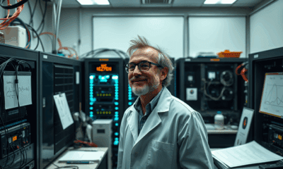
A Game-Changing Device for TB Diagnoses: Revolutionizing Healthcare for Children
This handheld device is the first that can detect tuberculosis in saliva, in addition to blood and sputum samples, an important breakthrough for testing children and HIV patients, who struggle to produce sputum. The device was found to deliver rapid, accurate results in under an hour, offering a cost-effective and accessible solution for diagnosing TB in resource-limited areas.


Biochemistry Research
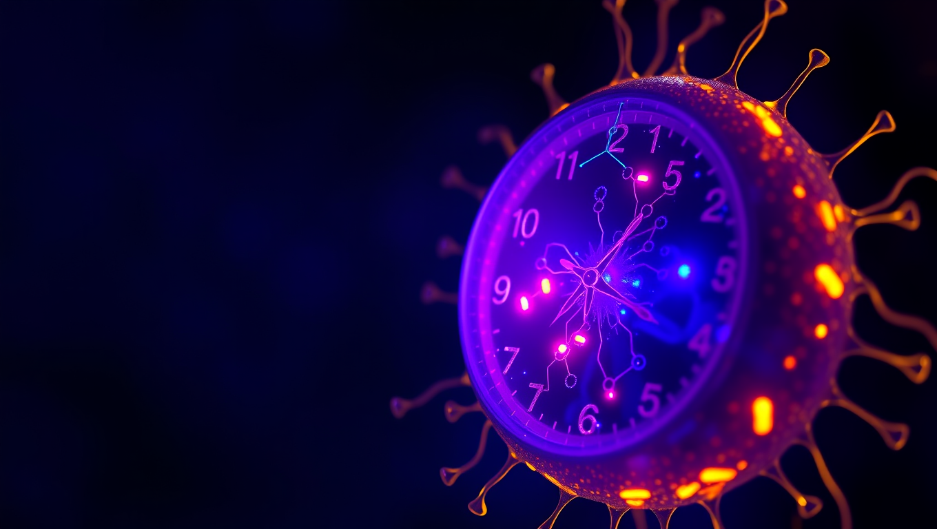
“Unlocking Timekeeping Secrets: Scientists Reveal How Artificial Cells Can Accurately Keep Rhythm”
Scientists at UC Merced have engineered artificial cells that can keep perfect time—mimicking the 24-hour biological clocks found in living organisms. By reconstructing circadian machinery inside tiny vesicles, the researchers showed that even simplified synthetic systems can glow with a daily rhythm—if they have enough of the right proteins.
Behavioral Science
The Sugar that Sparked Life: Unraveling the Mystery of Ribose’s Preeminence in RNA Development
What made ribose the sugar of choice for life’s code? Scientists at Scripps Research may have cracked a major part of this mystery. Their experiments show that ribose binds more readily and selectively to phosphate compared to other similar sugars, forming a structure ideal for RNA formation. This discovery hints at how nature might have selected specific molecules long before enzymes or life existed, and could reshape our understanding of life’s chemical origins.

Biochemistry Research
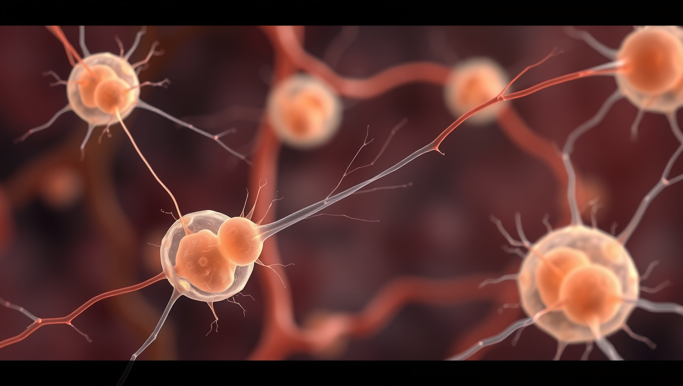
The Whispering Womb: Uncovering the Secret Language of Embryonic Cells
Scientists found that embryonic skin cells “whisper” through faint mechanical tugs, using the same force-sensing proteins that make our ears ultrasensitive. By syncing these micro-movements, the cells choreograph the embryo’s shape, a dance captured with AI-powered imaging and computer models. Blocking the cells’ ability to feel the whispers stalls development, hinting that life’s first instructions are mechanical. The discovery suggests hearing hijacked an ancient force-sensing toolkit originally meant for building bodies.

A New Horizon for Vision: How Gold Nanoparticles May Restore People’s Sight

Revolutionizing Quantum Communication: Direct Connections Between Multiple Processors

Retiring Abroad Can Be Lonely Business
Harnessing Water Waves: A Breakthrough in Controlling Floating Objects

Household Electricity Three Times More Expensive Than Upcoming ‘Eco-Friendly’ Aviation E-Fuels, Study Reveals

“Unveiling Hidden Patterns: A New Twist on Interference Phenomena”
Reducing Falls Among Elderly Women with Polypharmacy through Exercise Intervention
-
 Detectors1 year ago
A New Horizon for Vision: How Gold Nanoparticles May Restore People’s Sight
-
Cancer1 year ago
Revolutionizing Quantum Communication: Direct Connections Between Multiple Processors
-
Earth & Climate1 year ago
Retiring Abroad Can Be Lonely Business
-
Albert Einstein1 year ago
Harnessing Water Waves: A Breakthrough in Controlling Floating Objects
-
Earth & Climate1 year ago
Household Electricity Three Times More Expensive Than Upcoming ‘Eco-Friendly’ Aviation E-Fuels, Study Reveals
-
Chemistry1 year ago
“Unveiling Hidden Patterns: A New Twist on Interference Phenomena”
-
Diseases and Conditions1 year ago
Reducing Falls Among Elderly Women with Polypharmacy through Exercise Intervention
-
Agriculture and Food1 year ago
“A Sustainable Solution: Researchers Create Hybrid Cheese with 25% Pea Protein”


