
While we try to keep things accurate, this content is part of an ongoing experiment and may not always be reliable.
Please double-check important details — we’re not responsible for how the information is used.
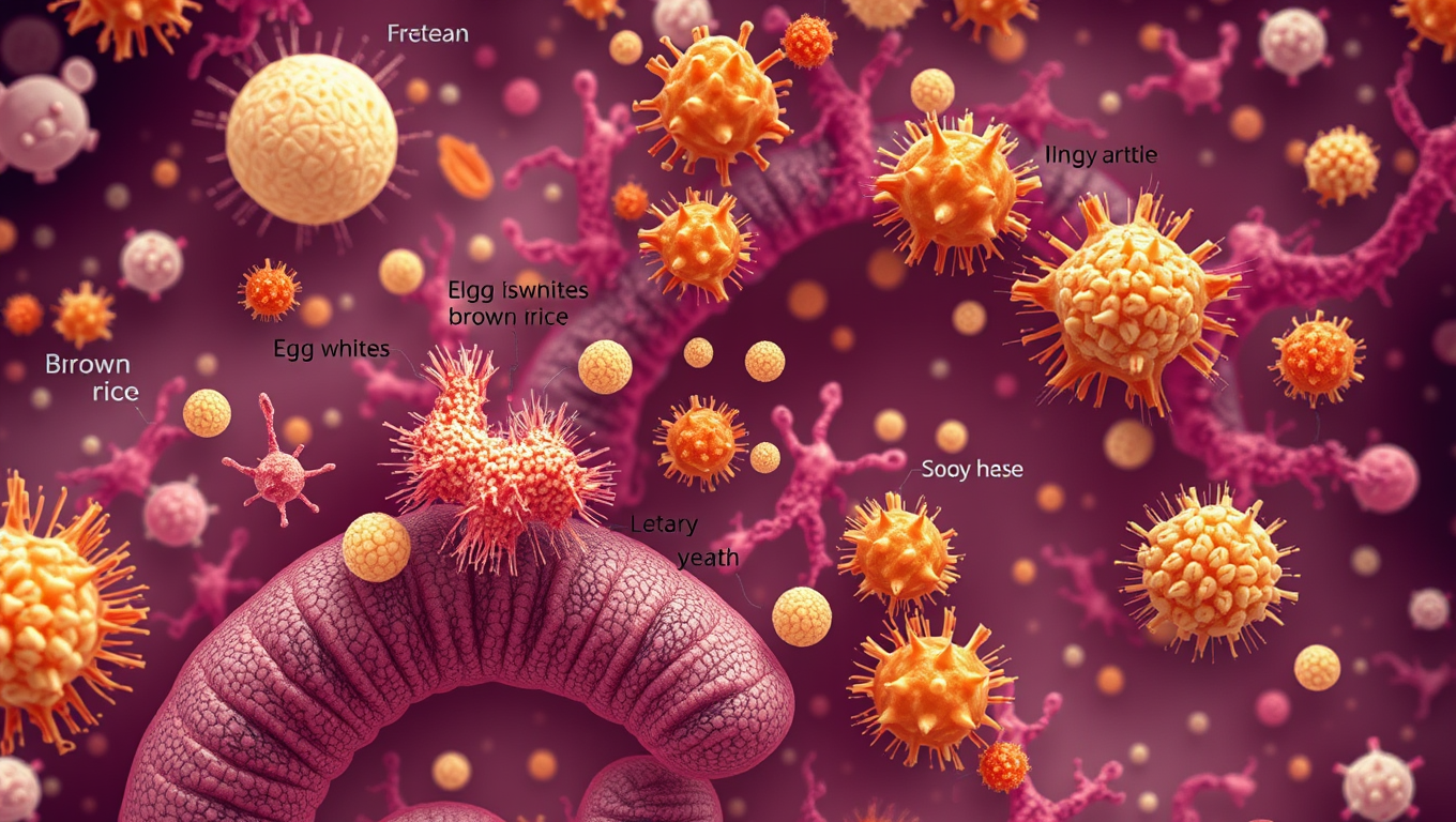
Cell Biology
“Protein Powerhouses: How Different Protein Sources Shape the Gut Microbiome”
Protein sources appear to have major effects on both the population and function of the mouse gut microbiome.


Behavioral Science
The Sugar that Sparked Life: Unraveling the Mystery of Ribose’s Preeminence in RNA Development
What made ribose the sugar of choice for life’s code? Scientists at Scripps Research may have cracked a major part of this mystery. Their experiments show that ribose binds more readily and selectively to phosphate compared to other similar sugars, forming a structure ideal for RNA formation. This discovery hints at how nature might have selected specific molecules long before enzymes or life existed, and could reshape our understanding of life’s chemical origins.

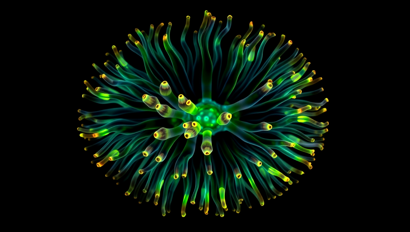
Cell Biology
A 600-Million-Year-Old Body Blueprint Uncovered in Sea Anemones
Sea anemones may hold the key to the ancient origins of body symmetry. A study from the University of Vienna shows they use a molecular mechanism known as BMP shuttling, once thought unique to bilaterally symmetrical animals like humans, insects, and worms. This surprising discovery implies that the blueprint for forming a back-to-belly body axis could date back over 600 million years, to a common ancestor of cnidarians and bilaterians.
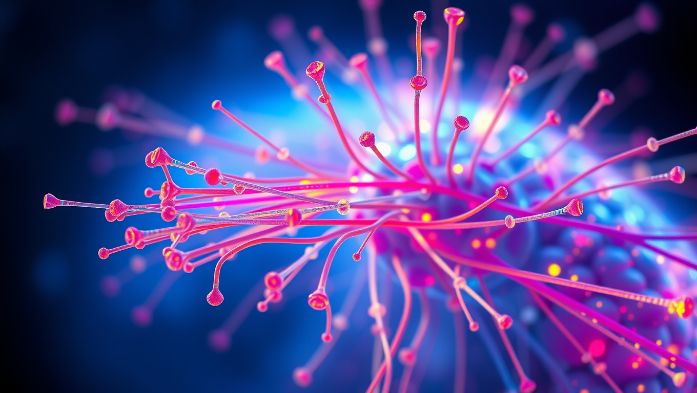
Biology
Unraveling Microtubule Mysteries: Scientists Crack Code on Cellular Scaffolding Secrets
Scientists found out how naturally unstable filaments decide whether to grow or to shorten.

A New Horizon for Vision: How Gold Nanoparticles May Restore People’s Sight

Revolutionizing Quantum Communication: Direct Connections Between Multiple Processors

Retiring Abroad Can Be Lonely Business
Harnessing Water Waves: A Breakthrough in Controlling Floating Objects

Household Electricity Three Times More Expensive Than Upcoming ‘Eco-Friendly’ Aviation E-Fuels, Study Reveals

“Unveiling Hidden Patterns: A New Twist on Interference Phenomena”
Reducing Falls Among Elderly Women with Polypharmacy through Exercise Intervention
-
 Detectors1 year ago
A New Horizon for Vision: How Gold Nanoparticles May Restore People’s Sight
-
Cancer1 year ago
Revolutionizing Quantum Communication: Direct Connections Between Multiple Processors
-
Earth & Climate1 year ago
Retiring Abroad Can Be Lonely Business
-
Albert Einstein1 year ago
Harnessing Water Waves: A Breakthrough in Controlling Floating Objects
-
Earth & Climate1 year ago
Household Electricity Three Times More Expensive Than Upcoming ‘Eco-Friendly’ Aviation E-Fuels, Study Reveals
-
Chemistry1 year ago
“Unveiling Hidden Patterns: A New Twist on Interference Phenomena”
-
Diseases and Conditions1 year ago
Reducing Falls Among Elderly Women with Polypharmacy through Exercise Intervention
-
Agriculture and Food1 year ago
“A Sustainable Solution: Researchers Create Hybrid Cheese with 25% Pea Protein”


