While we try to keep things accurate, this content is part of an ongoing experiment and may not always be reliable.
Please double-check important details — we’re not responsible for how the information is used.
Biochemistry
A Breakthrough in Brain Research: The Iontronic Pipette Revolutionizes Neurological Studies
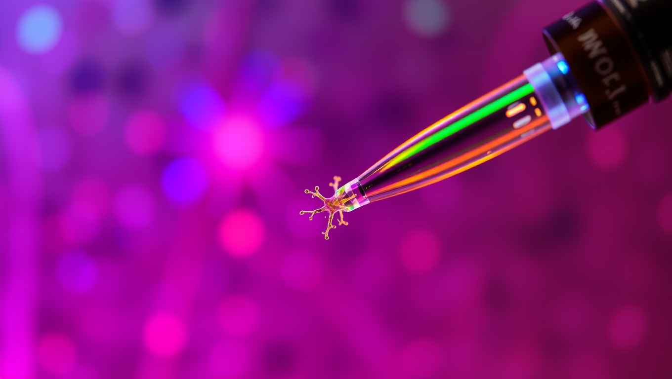
Researchers have developed a new type of pipette that can deliver ions to individual neurons without affecting the sensitive extracellular milieu. Controlling the concentration of different ions can provide important insights into how individual brain cells are affected, and how cells work together. The pipette could also be used for treatments.


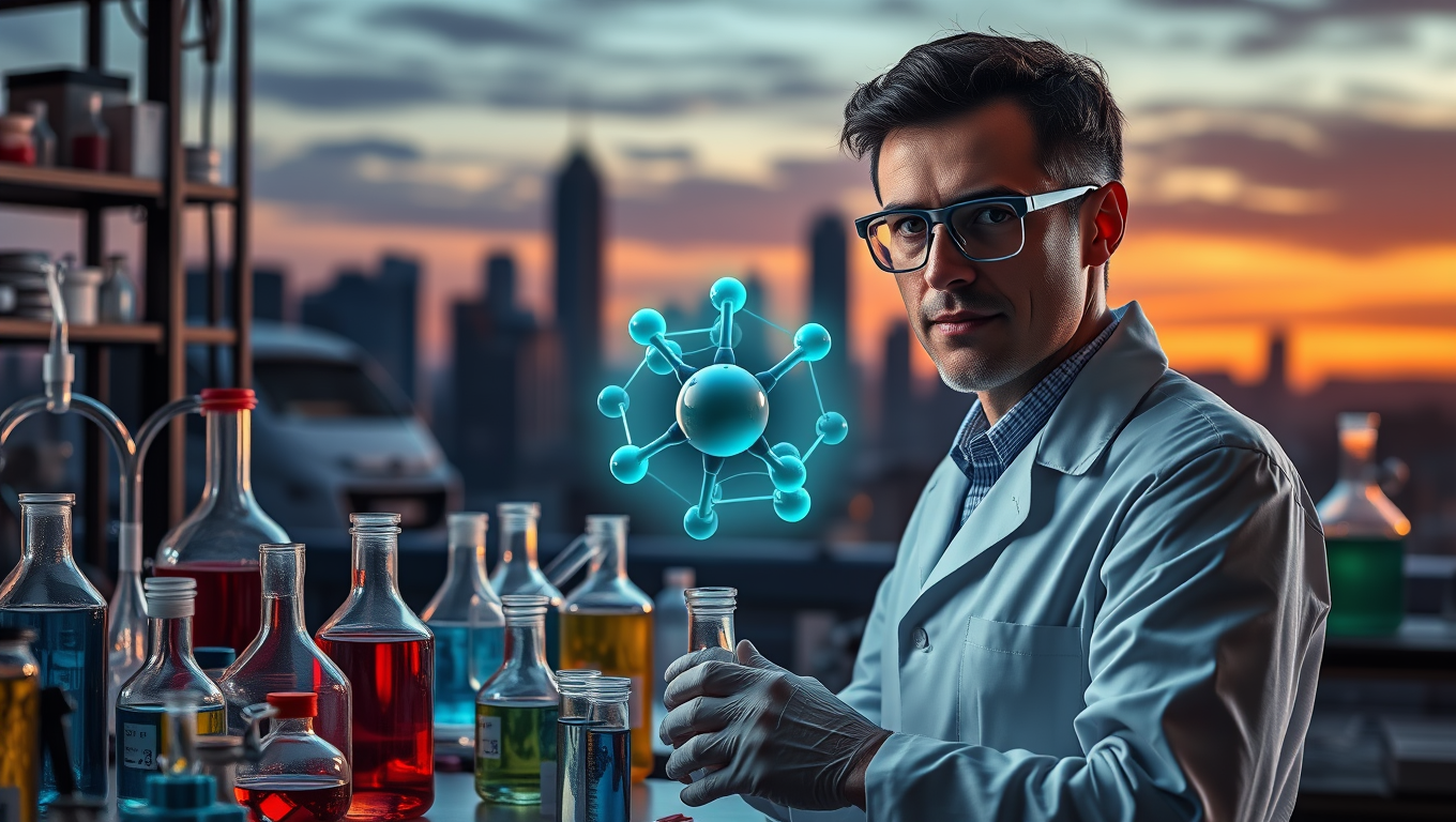
Biochemistry
Shape-Shifting Catalysts: Revolutionizing Green Chemistry with a Single Atom
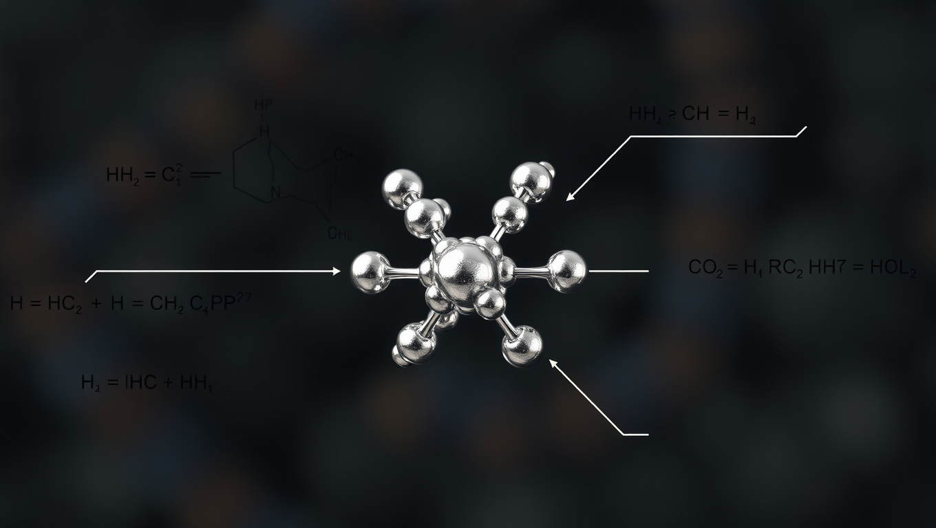
A team in Milan has developed a first-of-its-kind single-atom catalyst that acts like a molecular switch, enabling cleaner, more adaptable chemical reactions. Stable, recyclable, and eco-friendly, it marks a major step toward programmable sustainable chemistry.
Biochemistry
Scientists Finally Tame the Impossible: A Stable 48-Atom Carbon Ring is Achieved
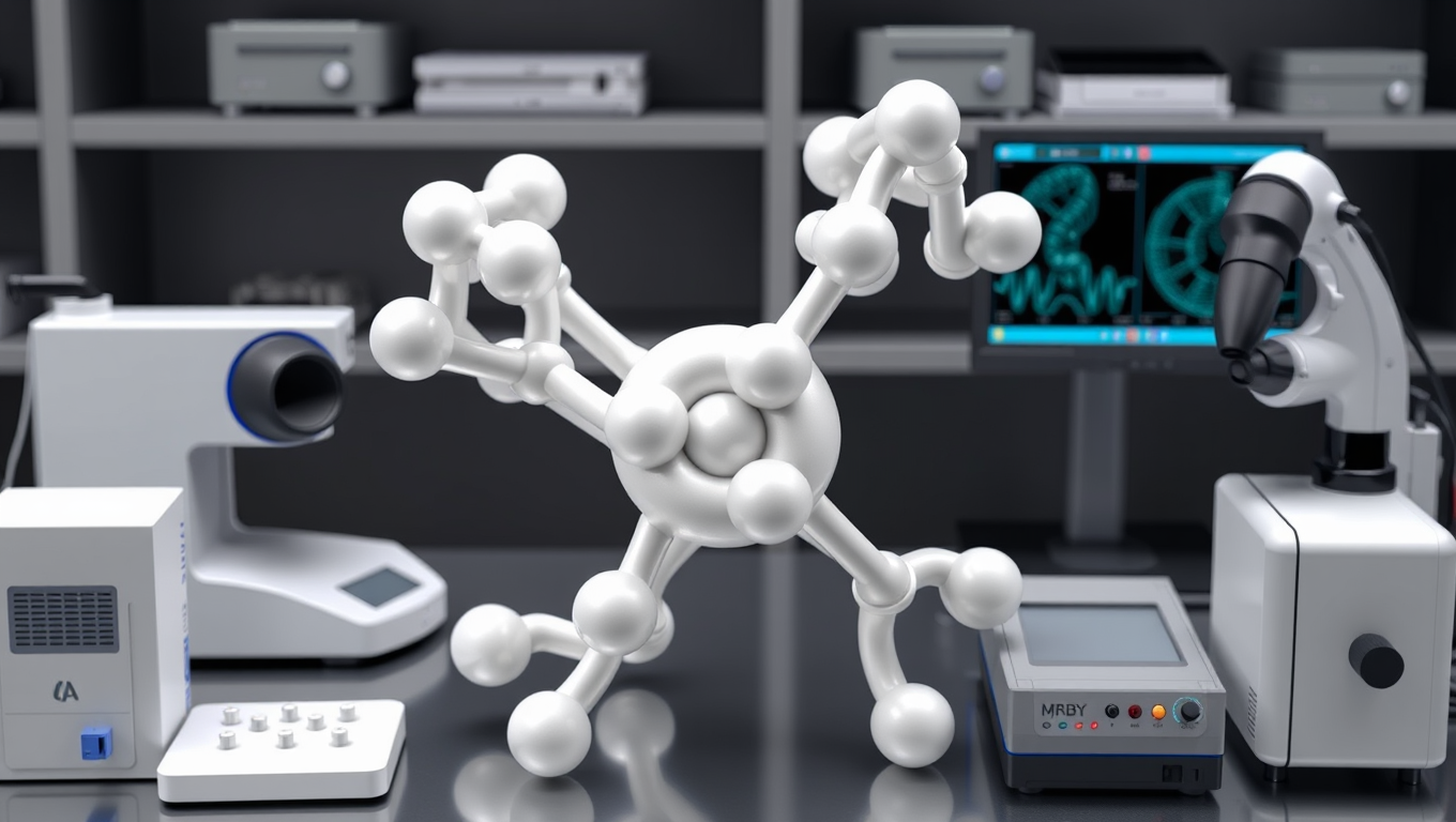
Researchers have synthesized a stable cyclo[48]carbon, a unique 48-carbon ring that can be studied in solution at room temperature, a feat never achieved before.
Biochemistry
“Revolutionizing Medicine: A 100x Faster Path to Life-Saving Drugs with Metal Carbenes”
Using a clever combo of iron and radical chemistry, scientists have unlocked a safer, faster way to create carbenes molecular powerhouses key to modern medicine and materials. It s 100x more efficient than previous methods.

A New Horizon for Vision: How Gold Nanoparticles May Restore People’s Sight

Revolutionizing Quantum Communication: Direct Connections Between Multiple Processors

Retiring Abroad Can Be Lonely Business

“Unveiling Hidden Patterns: A New Twist on Interference Phenomena”
Harnessing Water Waves: A Breakthrough in Controlling Floating Objects

Household Electricity Three Times More Expensive Than Upcoming ‘Eco-Friendly’ Aviation E-Fuels, Study Reveals
Reducing Falls Among Elderly Women with Polypharmacy through Exercise Intervention
-
 Detectors1 year ago
A New Horizon for Vision: How Gold Nanoparticles May Restore People’s Sight
-
Cancer1 year ago
Revolutionizing Quantum Communication: Direct Connections Between Multiple Processors
-
Earth & Climate1 year ago
Retiring Abroad Can Be Lonely Business
-
Chemistry1 year ago
“Unveiling Hidden Patterns: A New Twist on Interference Phenomena”
-
Albert Einstein1 year ago
Harnessing Water Waves: A Breakthrough in Controlling Floating Objects
-
Earth & Climate1 year ago
Household Electricity Three Times More Expensive Than Upcoming ‘Eco-Friendly’ Aviation E-Fuels, Study Reveals
-
Diseases and Conditions1 year ago
Reducing Falls Among Elderly Women with Polypharmacy through Exercise Intervention
-
Acid Rain1 year ago
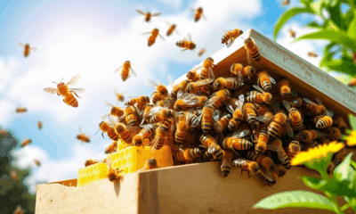
“Revolutionizing Honey Bee Survival: A New Pollen-Replacing Food Source Brings Hope for the Future”