Connect with us
We’re experimenting with AI-generated content to help deliver information faster and more efficiently.
While we try to keep things accurate, this content is part of an ongoing experiment and may not always be reliable.
Please double-check important details — we’re not responsible for how the information is used.

Cosmetic Surgery

“Microbes on Our Skin: The Hidden Heroes Against Sun Damage”

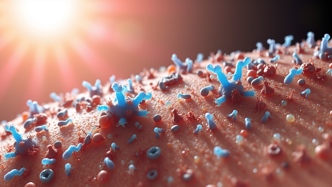
The skin microbiome plays an important role in health and disease. Researchers have now substantiated that certain skin bacteria can protect us from the sun’s ultraviolet (UV) radiation specifically by metabolizing cis-urocanic acid using an enzyme called urocanase. This enables the skin’s ability to fine-tune how it responds to UV radiation. The findings demonstrate the ability of the skin microbiome to remodel host immune functions.
Avatar photo

Published

on

The article you provided has been rewritten to improve clarity, structure, and style while maintaining its core ideas. Here’s the rewritten content:

Microbes on Our Skin: The Hidden Heroes Against Sun Damage

Researchers have made a groundbreaking discovery about the skin microbiome, revealing that certain bacteria can protect us from the bad effects of sunlight by metabolizing cis-urocanic acid using an enzyme called urocanase. This enables our skin to fine-tune its response to UV radiation.

The study, published in the Journal of Investigative Dermatology, demonstrates the ability of the skin microbiome to remodel host immune functions. Lead investigator VijayKumar Patra, PhD, explains that many internal and external factors influence the composition of the skin microbiome, including individual parameters such as race, gender, age, hormone levels, diet, and hygiene.

Researchers used a combination of microbiome sequencing, immunological assays, in vitro cultures, and gnotobiotic mouse models to study how skin bacteria respond to UVB radiation. They discovered that certain skin bacteria specifically metabolize cis-urocanic acid, a photoproduct of a major UV-absorbing chromophore of the stratum corneum, using an enzyme called urocanase.

Co-investigator Marc Vocanson, PhD, notes that this is the first time a direct metabolic link between UV radiation, a host-derived molecule, and bacterial behavior affecting immune function has been demonstrated. As interest grows in microbiome research and personalized medicine, understanding these microbe-host interactions could reshape the way we think about sun protection, immune diseases, skin cancer, or even treatments like phototherapy.

Co-investigator Peter Wolf, MD, concludes that these findings open the door to microbiome-aware sun protection, where we not only protect the skin from UV radiation but also consider how resident microbes can alter the immune landscape after exposure. In the future, topical treatments that modulate microbial metabolism could be used to minimize, maintain, or enhance UV-induced immunosuppression when clinically beneficial.

Noted expert Anna Di Nardo, MD, PhD, comments on the findings, saying that this pivotal study shows that microbial communities are not passive victims of environmental stress but dynamic regulators of immune responses. This newly uncovered role of microbial metabolism in modulating UV tolerance reshapes our understanding of the skin barrier – not just as a structural shield but as a metabolically active, microbially regulated interface.

With increasing concerns about UV exposure, skin aging, and cancer, a deeper understanding of this axis offers promising avenues for therapy and prevention.

Cosmetic Surgery

Unlocking the Innate Immune System: A New Path to Prevent Organ Transplant Rejection

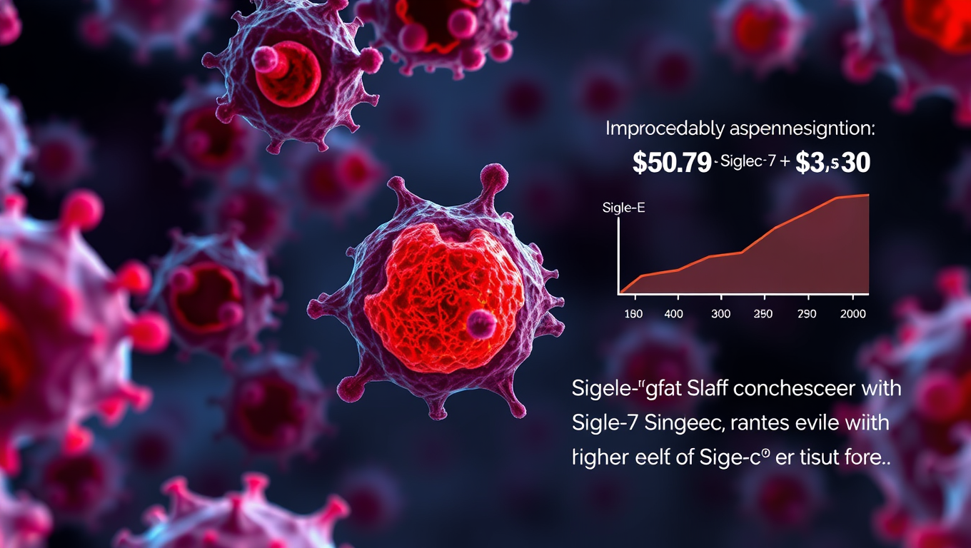
Researchers identified a natural ‘brake’ within the innate immune system: the inhibitory receptor Siglec-E (SigE) and its human counterparts, Siglec-7 and Siglec-9. This receptor helps prevent overactivation of immune cells that drive rejection. When this brake is missing, inflammation worsens, leading to faster rejection in preclinical models. Importantly, transplant patients with higher levels of Siglec-7 and Siglec-9 showed better graft survival, highlighting this pathway as a promising target for new therapies.

Avatar photo

Published

on

For decades, medical researchers have been searching for ways to prevent organ transplant rejection. Current treatments focus on suppressing T cells, part of the adaptive immune system. However, this approach has its limitations. A new study from Mass General Brigham sheds light on a previously untapped area: the innate immune system. Researchers identified a natural “brake” within this system – the inhibitory receptor Siglec-E (SigE) and its human counterparts, Siglec-7 and Siglec-9. This receptor plays a crucial role in preventing overactivation of immune cells that drive rejection.

When this brake is missing or malfunctioning, inflammation worsens, leading to faster rejection in preclinical models. Importantly, transplant patients with higher levels of Siglec-7 and Siglec-9 showed better graft survival, highlighting this pathway as a promising target for new therapies. Results were published in Science Translational Medicine.

“For decades, we’ve focused almost exclusively on controlling T cells to prevent rejection,” said Leonardo Riella, MD, PhD, medical director of Kidney Transplantation at Massachusetts General Hospital (MGH). “Our research shows that the innate immune system plays a pivotal role. By harnessing natural inhibitory pathways like Siglec-E, we can develop safer, more precise therapies that protect transplanted organs without compromising overall immune health.”

To conduct their studies, the researchers used mouse models of heart, kidney, and skin transplantation to study the roles of SigE. They found that recipients deficient in SigE had accelerated acute rejection and increased inflammation. The researchers also looked at the levels of the receptors in samples from human transplant biopsies, finding that higher levels of the receptors were associated with improved allograft survival.

“This discovery paves the way for next-generation treatments that address both arms of the immune system, offering hope for longer-lasting transplant success and reducing the need for lifelong immunosuppression,” said Riella.

Continue Reading

Biochemistry

Revolutionizing Skin Research with Artificial Hydrogels

Growing cells in the laboratory is an art that humans have mastered decades ago. Recreating entire three-dimensional tissues is much more challenging. Researchers are developing a new hydrogel-based material that makes it possible to engineer artificial skin tissues, which can serve as living three-dimensional models of human skin for better understanding and treating skin diseases.

Avatar photo

Published

on

Revolutionizing Skin Research with Artificial Hydrogels

The human skin is the largest organ, accounting for about 15% of our body weight. It plays a vital role in protecting us from pathogens, dehydration, and temperature extremes. However, skin diseases such as skin cancer, chronic wounds, and autoimmune skin diseases are widespread, yet often poorly understood.

To address this knowledge gap, researchers at Empa have been working on developing an artificial model of human skin. This living “artificial skin” will enable scientists to simulate skin diseases, ultimately leading to a better understanding of their causes and treatment options. Unlike computer or plastic models, this innovative approach involves creating a model that contains cells and emulates the layered and wrinkled structure of human skin.

To recreate such complexity, researchers require suitable building materials. Recently, Empa scientists have made significant progress in developing hydrogels that meet these complex requirements while being easy to manufacture. The basis of their success lies in gelatin derived from the skin of cold-water fish.

Hydrogels: A Suitable Substitute for Skin Extracellular Matrix

Like most tissues, human skin consists of cells embedded in a network of proteins and other biomolecules called extracellular matrix. This matrix provides the tissue with shape and structure while sustaining the cells. However, it differs from one tissue to another – even within different layers of the skin.

Researchers have discovered that hydrogels can simulate this complex extracellular matrix. These special polymers absorb large quantities of water and other fluids, making them ideal for recreating the skin’s extracellular matrix. Moreover, many hydrogels can be processed using a 3D printer, allowing researchers to combine multiple materials and cell types into a single structure – just like real skin.

The Empa team has developed a fish gelatin-based hydrogel that can be cross-linked in just a few steps. This non-swelling hydrogel can be printed with skin cells, enabling the creation of a 3D-printed artificial skin model that closely resembles human skin.

A Promising Tool for Wound Healing

The researchers’ innovative hydrogel has far-reaching implications. Without the addition of living cells, it can be used as a dressing material. This biologically compatible material is safer and carries a lower risk of disease transmission compared to materials based on mammalian gelatin.

Moreover, the fish skin from which this hydrogel is derived is being researched as a promising tool for wound healing. The Empa team’s hydrogel is more homogeneous and can be tailored precisely to meet patient needs – with different shapes, thicknesses, and firmness levels.

The researchers have applied for a patent for their fish gelatin-based hydrogel and plan to finish developing the living skin model. This will make it available to other scientists, promoting a better understanding of skin disease development and treatment options.

Continue Reading

Asthma

Predicting Surgery Success with Chronic Rhinosinusitis – A New Risk Score

A new CT-scan based risk score facilitates the identification of patients at risk of revision endoscopic sinus surgery due to chronic rhinosinusitis.

Avatar photo

Published

on

Chronic rhinosinusitis, a condition characterized by inflammation of the nasal and sinus cavities, affects millions of people worldwide. Symptoms include nasal congestion, mucus secretion, and pressure in the sinuses. In severe cases, the disease can be associated with asthma or NSAID-exacerbated respiratory disease (N-ERD). The standard treatment involves nasally administered corticosteroids, which can be supplemented with orally administered corticosteroids for the polypoid form of the disease.

For patients who do not respond to medical treatments, endoscopic sinus surgery is often considered. However, a significant number of patients may require revision surgery due to persistent symptoms and disease progression. In fact, research suggests that up to 28% of patients undergoing endoscopic sinus surgery for chronic rhinosinusitis may need revision surgery within one year.

A new study published in Clinical and Translational Allergy has introduced a novel CT-scan based risk score to predict the likelihood of revision endoscopic sinus surgery. The Sinonasal Radiological Score (SR score) takes into account indicators such as non-detectable anatomy of the nasal turbinates, which is often associated with polypoid mucosal swelling, and obstructed drainage of the frontal sinus.

In comparison to the existing Lund-Mackay scoring system, the SR score provides a more accurate assessment of the risk of revision surgery. The study analyzed data from 483 patients with chronic rhinosinusitis, including those who underwent endoscopic sinus surgery within one year following their CT scan. Results showed that conditions such as asthma and N-ERD increased the risk of revision surgery.

This breakthrough research highlights the importance of early prediction of disease progression and planning for further treatment. By using the new SR score in conjunction with conventional medical and surgical treatments, clinicians can identify patients at risk of revision surgery and tailor their treatment approach accordingly.

Continue Reading

Trending