Connect with us
We’re experimenting with AI-generated content to help deliver information faster and more efficiently.
While we try to keep things accurate, this content is part of an ongoing experiment and may not always be reliable.
Please double-check important details — we’re not responsible for how the information is used.

Depression

Decoding Pain’s Dark Side: Uncovering a Hidden Brain Circuit Behind Fibromyalgia, Migraines, and PTSD

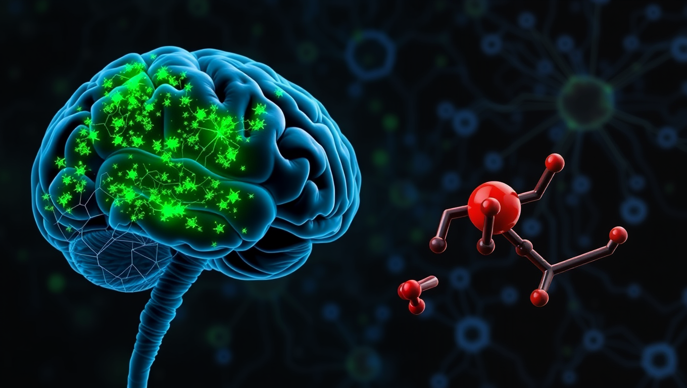
What if your brain is the reason some pain feels unbearable? Scientists at the Salk Institute have discovered a hidden brain circuit that gives pain its emotional punch—essentially transforming ordinary discomfort into lasting misery. This breakthrough sheds light on why some people suffer more intensely than others from conditions like fibromyalgia, migraines, and PTSD. By identifying the exact group of neurons that link physical pain to emotional suffering, the researchers may have found a new target for treating chronic pain—without relying on addictive medications.
Avatar photo

Published

on

The way we perceive and respond to physical pain is more than just a physical sensation – it also carries an emotional weight. This emotional discomfort can motivate us to take action and helps us learn to associate negative feelings with situations so we can avoid them in the future. However, when our ability to tolerate pain becomes too sensitive or lasts too long, it can result in chronic and affective pain disorders such as fibromyalgia, migraines, and post-traumatic stress disorder (PTSD).

Researchers at the Salk Institute have now identified a brain circuit that gives physical pain its emotional tone. Published in the Proceedings of the National Academy of Sciences, the study reveals a group of neurons in the central brain area called the thalamus that appears to mediate the emotional or affective side of pain in mice.

The prevailing view for decades was that the brain processes sensory and emotional aspects of pain through separate pathways. However, this new pathway challenges the textbook understanding of how pain is processed in the brain and body. The physical sensation of pain allows you to immediately detect it, assess its intensity, and identify its source, while the affective part of pain makes it unpleasant.

This distinction is crucial because most people start to perceive pain at the same stimulus intensities. However, our ability to tolerate pain varies greatly, with some individuals being more sensitive than others. The affective processing determines how much we suffer or feel threatened by pain. If this becomes too sensitive or lasts too long, it can result in a pain disorder.

The researchers used advanced techniques to manipulate the activity of specific brain cells and discovered a new spinothalamic pathway in mice. In this circuit, pain signals are sent from the spinal cord into a different part of the thalamus, which has connections to the amygdala, the brain’s emotional processing center. This particular group of neurons can be identified by their expression of CGRP (calcitonin gene-related peptide).

When these CGRP neurons were “turned off,” the mice still reacted to mild pain stimuli but didn’t seem to associate lasting negative feelings with these situations. However, when these same neurons were “turned on,” the mice showed clear signs of distress and learned to avoid that area, even when no pain stimuli had been used.

“Pain processing is not just about nerves detecting pain; it’s about the brain deciding how much that pain matters,” says first author Sukjae Kang. Understanding the biology behind these two distinct processes will help us find treatments for the kinds of pain that don’t respond to traditional drugs.

Many chronic pain conditions, such as fibromyalgia and migraine, involve long, intense, unpleasant experiences of pain often without a clear physical source or injury. Some patients also report extreme sensitivity to ordinary stimuli like light, sound, or touch which others would not perceive as painful.

Han says overactivation of the CGRP spinothalamic pathway may contribute to these conditions by making the brain misinterpret or overreact to sensory inputs. In fact, transcriptomic analysis of the CGRP neurons showed that they express many of the genes associated with migraine and other pain disorders.

Several CGRP blockers are already being used to treat migraines. This study may help explain why these medications work and could inspire new nonaddictive treatments for affective pain disorders. Han also sees potential relevance for psychiatric conditions that involve heightened threat perception, such as PTSD. Quieting this pathway with CGRP blockers could offer a new approach to easing fear, avoidance, and hypervigilance in trauma-related disorders.

Importantly, the relationship between the CGRP pathway and the psychological pain associated with social experiences like grief, loneliness, and heartbreak remains unclear and requires further study.

“Our discovery of the CGRP affective pain pathway gives us a molecular and circuit-level explanation for the difference between detecting physical pain and suffering from it,” says Han. “We’re excited to continue exploring this pathway and enabling future therapies that can reduce this suffering.”

Depression

Uncovering the Link Between Sensitivity and Mental Health Conditions

Researchers analyzing 33 studies found strong evidence that highly sensitive people are more prone to depression and anxiety but also more likely to benefit from therapy. Since about 31% of the population is highly sensitive, experts argue that clinicians should consider sensitivity levels when diagnosing and treating mental health conditions.

Avatar photo

Published

on

Uncovering the Link Between Sensitivity and Mental Health Conditions

For decades, mental health studies have focused on neuroticism as a primary factor in diagnosing conditions like depression and anxiety. However, researchers from Queen Mary University and the University of Surrey have conducted the first meta-analysis to examine the relationship between sensitivity and common mental health problems. The study, which analyzed 33 studies, reveals a significant positive correlation between sensitivity and depression, anxiety, post-traumatic stress disorder (PTSD), agoraphobia, and avoidant personality disorder.

Sensitivity was defined as a personality trait that allows individuals to perceive and process environmental stimuli, such as subtle changes in their surroundings or the emotions of those around them. This often-overlooked trait is crucial for understanding an individual’s mental health, particularly when it comes to developing effective treatment plans.

Tom Falkenstein, a psychotherapist and PhD student at Queen Mary University, emphasized the importance of considering sensitivity in clinical practice: “Our findings suggest that sensitivity should be considered more in clinical practice. This could lead to improved diagnosis and treatment for individuals who are highly sensitive.”

Falkenstein also noted that around 31% of the general population are considered highly sensitive and may respond better to certain psychological interventions, such as applied relaxation and mindfulness techniques. By taking sensitivity into account, mental health professionals can tailor treatment plans to meet the unique needs of each individual.

Michael Pluess, Professor in Developmental Psychology at the University of Surrey and Visiting Professor at Queen Mary University, added: “Our results provide further evidence that sensitive people are more affected by both negative and positive experiences. This highlights the importance of a supportive environment for their well-being.”

The systematic review and meta-analysis were carried out by an academic team from several universities, including Queen Mary University and the University of Surrey.

The study’s findings have significant implications for mental health care professionals, who should now consider sensitivity when developing treatment plans for individuals with depression, anxiety, PTSD, agoraphobia, or avoidant personality disorder. By doing so, they can provide more effective and personalized care to those in need.

Continue Reading

Depression

Groundbreaking Discovery Offers Hope for PTSD Patients

Researchers discovered that PTSD may be driven by excess GABA from astrocytes, not neurons. This chemical imbalance disrupts the brain’s ability to forget fear. A new drug, KDS2010, reverses this effect in mice and is already in human trials. It could represent a game-changing therapy.

Avatar photo

Published

on

Groundbreaking Discovery Offers Hope for PTSD Patients

For decades, patients suffering from post-traumatic stress disorder (PTSD) have struggled to forget traumatic memories, even after the danger has passed. The failure to extinguish these fear memories has long puzzled scientists and presented a significant hurdle in treatment, particularly since current medications targeting serotonin receptors offer limited relief for only a subset of patients.

A recent discovery by researchers at the Institute for Basic Science (IBS) and Ewha Womans University has uncovered a new brain mechanism driving PTSD. Led by Dr. C. Justin LEE at the IBS Center for Cognition and Sociality and Professor LYOO In Kyoon at Ewha Womans University, the team found that excessive GABA produced by astrocytes impairs the brain’s ability to extinguish fear memories. This deficit is a core feature of PTSD, explaining why traumatic memories can persist long after the threat has passed.

The researchers discovered that a highly selective and reversible monoamine oxidase B (MAOB) inhibitor called KDS2010 can reverse PTSD-like symptoms in mice. The drug has already passed Phase 1 safety trials in humans, making it a strong candidate for future PTSD treatments.

PTSD remains challenging to treat, with current medications targeting serotonin pathways providing limited relief for many patients. The new study focused on the medial prefrontal cortex (mPFC), a region of the brain critical for regulating fear. Importantly, GABA levels decreased in patients who showed clinical improvement, highlighting the chemical’s central role in recovery.

To uncover the origin of this excess GABA, the researchers examined postmortem human brain tissue and used PTSD-like mouse models. They found that astrocytes were producing abnormal amounts of GABA via the enzyme monoamine oxidase B (MAOB). This astrocyte-derived GABA impaired neural activity, blocking the brain’s ability to forget traumatic memories.

When KDS2010 was administered to mice, they showed normalized brain activity and were able to extinguish fear responses. The drug reduced GABA levels, restored blood flow in the mPFC, and re-enabled memory extinction mechanisms. This study confirms astrocytic MAOB as a central driver of PTSD symptoms and MAOB inhibition as a viable therapeutic path.

A major challenge was linking clinical findings in humans with cellular mechanisms in the lab. The researchers applied a “reverse translational” strategy: they began with clinical brain scans, moved backward to identify the cellular source of dysfunction, then confirmed the mechanism and tested drug effects in animal models. This approach led to a new understanding of how glial cells actively shape psychiatric symptoms.

“This study is the first to identify astrocyte-derived GABA as a key pathological driver of fear extinction deficit in PTSD,” said Dr. WON Woojin, a postdoctoral researcher and co-first author of the study. “Our findings not only uncover a novel astrocyte-based mechanism underlying PTSD but also provide preclinical evidence for a new therapeutic approach using an MAOB inhibitor.”
Director C. Justin LEE emphasized that “This work represents a successful example of reverse translational research, where clinical findings in humans guided the discovery of underlying mechanisms in animal models. By identifying astrocytic GABA as a pathological driver in PTSD and targeting it via MAOB inhibition, the study opens a completely new therapeutic paradigm not only for PTSD but also for other neuropsychiatric disorders such as panic disorder, depression, and schizophrenia.”
The researchers plan to further investigate astrocyte-targeted therapies for various neuropsychiatric disorders. With KDS2010 currently undergoing Phase 2 clinical trials, this discovery may soon lead to new options for patients whose symptoms have not responded to conventional treatments.

Continue Reading

Depression

The Unseen Toll of the Pandemic: How Stress and Isolation May Be Aging Your Brain

Even people who never caught Covid-19 may have aged mentally faster during the pandemic, according to new brain scan research. This large UK study shows how the stress, isolation, and upheaval of lockdowns may have aged our brains, especially in older adults, men, and disadvantaged individuals. While infection itself impacted some thinking skills, even those who stayed virus-free showed signs of accelerated brain aging—possibly reversible. The study highlights how major life disruptions, not just illness, can reshape our mental health.

Avatar photo

Published

on

The COVID-19 pandemic has left an indelible mark on humanity, but its impact may go beyond the physical toll of the virus itself. A recent study suggests that even those who never contracted COVID-19 may have experienced accelerated brain aging due to the stresses and disruptions caused by the pandemic.

Led by experts at the University of Nottingham, the research team analyzed longitudinal brain scans from nearly 1,000 healthy adults, taken as part of the UK Biobank study. Some participants had scans before and after the pandemic, while others only had pre-pandemic scans. Using advanced imaging and machine learning, the researchers estimated each person’s “brain age” – how old their brain appeared to be compared to their actual age.

The findings were striking: people who lived through the pandemic showed signs of faster brain aging over time than those scanned entirely before it. The changes were most noticeable in older individuals, men, and those from more disadvantaged backgrounds. Notably, only participants who were infected with COVID-19 between their scans showed a drop in certain cognitive abilities, such as mental flexibility and processing speed.

“This study reminds us that brain health is shaped not only by illness but by our everyday environment,” said Dorothee Auer, Professor of Neuroimaging and senior author on the study. “The pandemic put a strain on people’s lives, especially those already facing disadvantage. We can’t yet test whether the changes we saw will reverse, but it’s certainly possible, and that’s an encouraging thought.”

Stress, isolation, and global disruption may have left their mark on people’s minds, leading to accelerated brain aging. However, the study highlights the possibility of reversibility, offering a glimmer of hope for those affected. The pandemic has taught us that even in the face of adversity, our brains can adapt and potentially recover – a testament to the resilience of the human spirit.

Continue Reading

Trending