While we try to keep things accurate, this content is part of an ongoing experiment and may not always be reliable.
Please double-check important details — we’re not responsible for how the information is used.
Animals
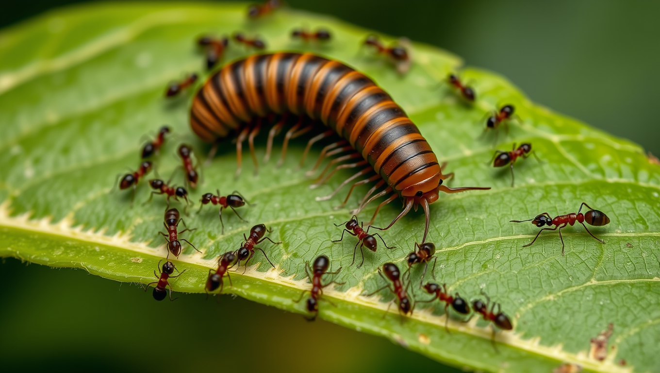
“From Millipede Secretions to Human Pain Relief — A New Path for Drug Discovery”
Millipedes, often dismissed as creepy crawlies, may hold the secret to future painkillers and neurological drugs. Researchers at Virginia Tech discovered unique alkaloid compounds in the defensive secretions of a native millipede species. These complex molecules, which cause disorientation in ants, interact with human neuroreceptors linked to pain and cognition. By decoding these natural chemical defenses, scientists could open a new path toward innovative drug therapies, though challenges remain in producing the compounds at scale.


Animals
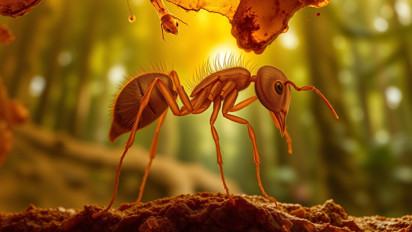
Unveiling the Ancient Secrets of the Dirt Ant: A 16-million-year-old Fossil Reveals the Smallest Predator Ant Ever Found
A fossilized Caribbean dirt ant, Basiceros enana, preserved in Dominican amber, reveals the species ancient range and overturns assumptions about its size evolution. Advanced imaging shows it already had the camouflage adaptations of modern relatives, offering new insights into extinction and survival strategies.
Animals
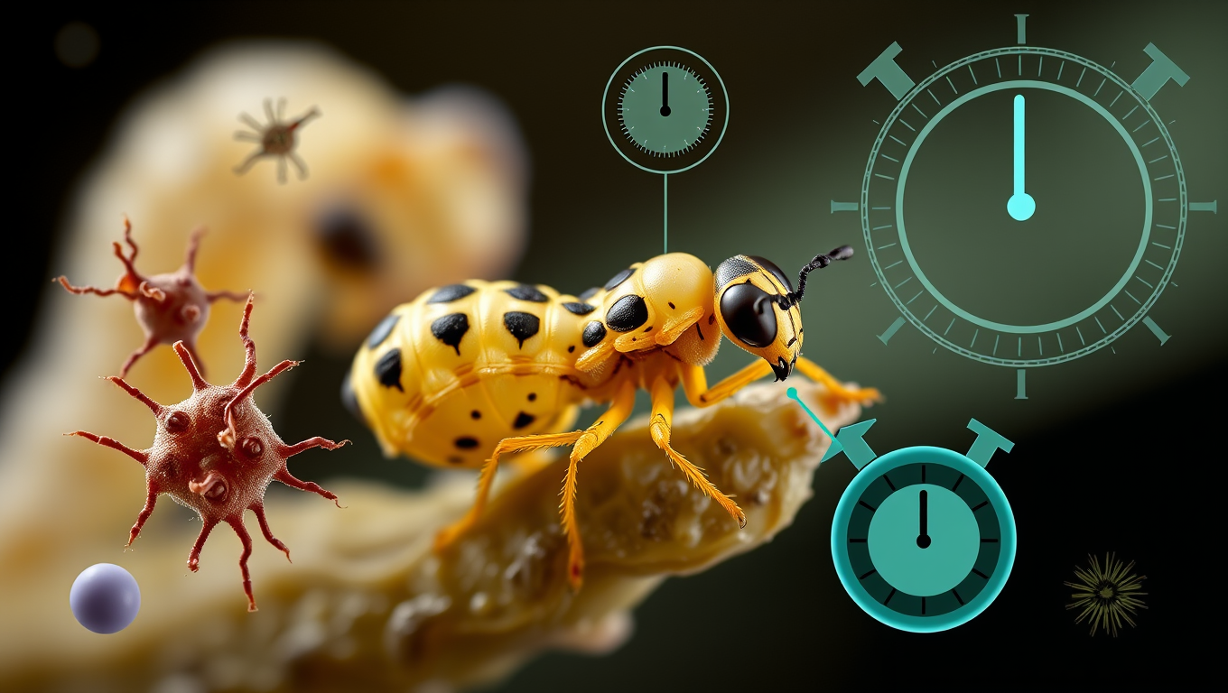
Nature’s Anti-Aging Hack? Jewel Wasp Larvae Slow Their Biological Clock
Scientists discovered that jewel wasp larvae that undergo a developmental “pause” live longer and age more slowly at the molecular level by nearly 30%. This slowdown is tied to conserved biological pathways, hinting at possible applications for human aging.
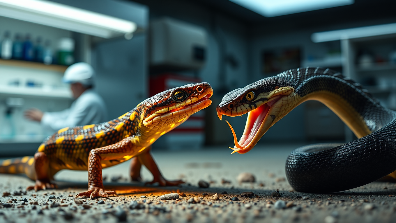
Animals
“Nature’s Armor: Scientists Uncover Gene Behind Aussie Skinks’ Immunity to Deadly Snake Venom”
Australian skinks have developed a remarkable genetic defense against venomous snake bites by mutating a key muscle receptor, making them resistant to neurotoxins. These tiny but powerful molecular changes mirror those found in cobra-resistant mammals like mongooses and honey badgers. This evolutionary arms race not only shows how adaptable life can be but also offers exciting possibilities for creating new antivenoms and therapies in human medicine.

A New Horizon for Vision: How Gold Nanoparticles May Restore People’s Sight

Retiring Abroad Can Be Lonely Business

Revolutionizing Quantum Communication: Direct Connections Between Multiple Processors

Household Electricity Three Times More Expensive Than Upcoming ‘Eco-Friendly’ Aviation E-Fuels, Study Reveals

“Unveiling Hidden Patterns: A New Twist on Interference Phenomena”
Harnessing Water Waves: A Breakthrough in Controlling Floating Objects
Reducing Falls Among Elderly Women with Polypharmacy through Exercise Intervention
-
 Detectors1 year ago
A New Horizon for Vision: How Gold Nanoparticles May Restore People’s Sight
-
Earth & Climate1 year ago
Retiring Abroad Can Be Lonely Business
-
Cancer1 year ago
Revolutionizing Quantum Communication: Direct Connections Between Multiple Processors
-
Earth & Climate1 year ago
Household Electricity Three Times More Expensive Than Upcoming ‘Eco-Friendly’ Aviation E-Fuels, Study Reveals
-
Chemistry1 year ago
“Unveiling Hidden Patterns: A New Twist on Interference Phenomena”
-
Albert Einstein1 year ago
Harnessing Water Waves: A Breakthrough in Controlling Floating Objects
-
Diseases and Conditions1 year ago
Reducing Falls Among Elderly Women with Polypharmacy through Exercise Intervention
-
Acid Rain1 year ago
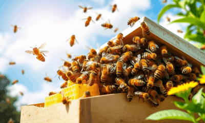
“Revolutionizing Honey Bee Survival: A New Pollen-Replacing Food Source Brings Hope for the Future”