Connect with us

Biochemistry Research

A Breakthrough in Viral Detection: Enhanced Sensitivity and Selectivity for Early Identification

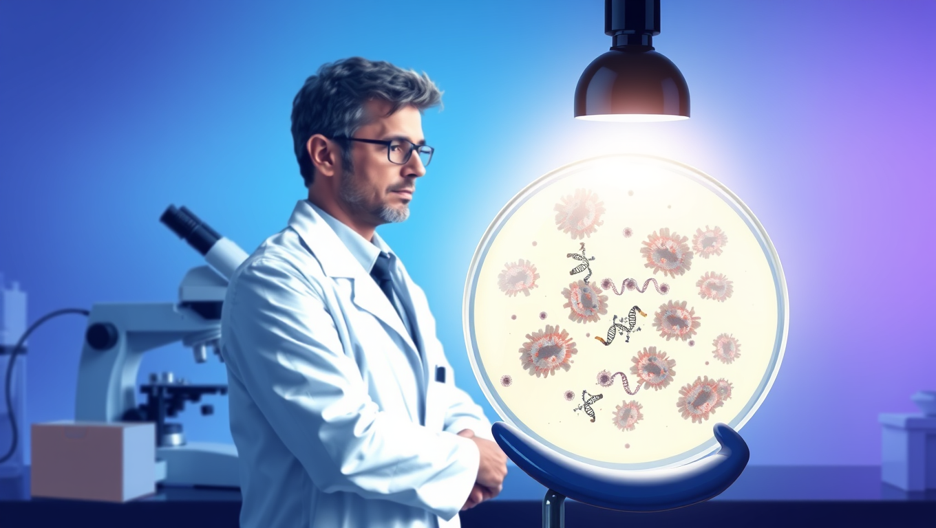
Researchers have unveiled innovative, label-free ratiometric fluorosensor designed for the selective and sensitive detection of enteroviral RNA. The research promises to deliver even more advanced and effective detection methods, reinforcing the importance of interdisciplinary collaboration in addressing global health challenges.
Avatar photo

Published

on

The rewritten article is as follows:

In a significant leap forward for viral research, scientists from the Nanoscience Center at the University of Jyväskylä, Finland, have developed an innovative, label-free ratiometric fluorosensor designed for the selective and sensitive detection of enteroviral RNA. This advancement promises to deliver even more effective detection methods, underscoring the importance of interdisciplinary collaboration in addressing global health challenges.

Viruses pose a severe threat to global health, as evidenced by recent pandemics. Early detection and identification are crucial for preventing new outbreaks. Traditional detection methods, while effective, often lack the possibility to provide spatiotemporal information on virus genome release.

“This interdisciplinary effort, combining expertise from biology, chemistry, and physics, marks a significant advancement in viral detection technology,” says Professor Jussi Toppari from the University of Jyväskylä. “We have developed an enhanced ratiometric fluorosensor using carbon dots (CDs) functionalized with Probe and ethidium bromide for detection of enteroviral RNA.”

The innovative ratiometric fluorosensor is a powerful tool for bioanalyte sensing, with CDs leading the way due to their exceptional properties. These unique characteristics position CDs as a game-changer in the field of biosensing.

“Our Functionalized Sensor (Func Sensor), where CDs are functionalized with the probe clearly outperforms the more traditional approach of Non-Functionalized Sensor (Non-Func Sensor),” explains Doctoral Researcher Amar Raj from University of Jyväskylä. “The Func Sensor showed a higher sensitivity with DNA and real viral RNA, exhibiting improved selectivity.”

The superior performance of the Func Sensor is attributed to enhanced charge transfer due to covalent functionalization.

“Our proof-of-principle study highlights the importance of covalent immobilization of the probe for improved electron transfer between CDs and EB,” says Professor Varpu Marjomäki from University of Jyväskylä. “This demonstrates the suitability of the Func sensor for practical applications in rapid, real-time, and precise in situ detection of viral RNA.”

The research also shows that the Func sensor can detect enteroviral RNA release from the capsid in real-time in vitro.

“This means that the Func sensor can be used as a novel viral RNA sensing platform offering a much-needed possibility to detect real-time viral RNA appearance during infection,” says Marjomäki.

Towards safer research, this pioneering work demonstrates a novel method for detecting viral RNA and sheds light on charge transfer mechanisms between fluorophores. Building on this success, the research team is now working to make the system more robust by replacing potentially hazardous dyes with less cytotoxic ones.

“This enhancement will further improve the safety and efficacy of in vivo viral RNA detection,” according to Pathak.

Biochemistry Research

Unveiling the Origins of HIV Resistance: A 7,000-Year-Old Genetic Mutation Reveals Ancient Secrets

Modern HIV medicine is based on a common genetic mutation. Now, researchers have traced where and when the mutation arose — and how it protected our ancestors from ancient diseases.

Avatar photo

Published

on


The story begins with a simple yet fascinating connection between modern HIV medicine and an ancient human from the Black Sea region. Researchers at the University of Copenhagen have now unraveled the mystery behind a millennia-old genetic mutation that protects against HIV, affecting 18-25% of the Danish population. This breakthrough has shed light on the origins of this crucial genetic variation.

The researchers employed advanced DNA technology to analyze the genetic material of over 2,000 living people worldwide and developed an AI-based method to identify the mutation in ancient DNA from old bones. By examining data from over 900 skeletons dating from the early Stone Age to the Viking Age, they pinpointed the region where the mutation originated – a person from the Black Sea region up to 9,000 years ago.

But why did this genetic mutation arise and spread rapidly among our ancestors? The researchers believe it provided an advantage in surviving during a time when humans were exposed to new pathogens. This variation disrupted an immune gene, which may have been beneficial by dampening the immune system. As humans transitioned from hunter-gatherers to living closely together in agricultural societies, the pressure from infectious diseases increased, and a more balanced immune system may have been advantageous.

The discovery of this 7,000-year-old genetic mutation not only reveals ancient secrets but also provides valuable insights into modern HIV medicine. It highlights the importance of understanding our evolutionary history and how it has shaped our genetic makeup. This breakthrough opens up new avenues for research, potentially leading to innovative treatments for various diseases.

Continue Reading

Biochemistry Research

A More Realistic Look at DNA in Action: Researchers Shed Light on the Mechanical Forces Behind Strand Separation

By creating a more true-to-life representation of DNA’s environment, researchers have discovered that strand separation may take more mechanical force than the field previously believed.

Avatar photo

Published

on

The way we study DNA has long been a topic of discussion among researchers. Traditionally, biochemistry labs isolate DNA within a water-based solution that allows scientists to manipulate it without interacting with other molecules. However, this approach can be misleading, as it doesn’t reflect the true environment of a living cell. In fact, the interior of a cell is “super crowded” with molecules, which can significantly impact the behavior of DNA.

Researchers at Northwestern University have taken a more realistic approach to studying DNA by creating an environment that mimics the conditions within a living cell. Led by Professor John Marko, the team used microscopic magnetic tweezers to separate DNA and then carefully attach strands of it to surfaces on one end, and tiny magnetic particles on the other. This allowed them to conduct high-tech imaging and investigate how different types of molecules interact with DNA.

The researchers found that strand separation, a crucial process for initiating replication or making repairs, may require more mechanical force than previously believed. They introduced three types of molecules to the solution holding DNA, mimicking proteins and investigating interactions among glycerol, ethylene glycol, and polyethylene glycol (each approximately the size of one DNA double helix, two or three nanometers).

“We wanted to have a wide variety of molecules where some cause dehydration, destabilizing DNA mechanically, and then others that stabilize DNA,” said Northwestern post-doctoral researcher Parth Desai. “It’s not exactly analogous to things found in cells, but you could imagine that other competing proteins in cells will have a similar effect.”
The team wrote a paper on their findings, which will be published on June 17 in the Biophysical Journal. Marko and Desai hope to run more experiments that incorporate multiple crowding agents and move closer to a true representation of a cell.

“If this affects DNA strand separation, all protein interactions with DNA are also going to be affected,” said Marko. “For example, the tendency for proteins to stick to specific sites on DNA and to control specific processes — this is also going to be altered by crowding.”

Their research has significant implications for understanding fundamental biochemical processes and may lead to new medical advances. The team hopes to study how interactions between enzymes and DNA are impacted by crowding in a living cell, which could have far-reaching consequences for our understanding of cellular biology.

This work was supported by the National Institutes of Health (grant R01-GM105847) and by subcontract to the University of Massachusetts Center for 3D Structure and Physics of the Genome (under NIH grant UM1-HG011536).

Continue Reading

Bacteria

Unlocking Efficiency: Researchers Reveal Secrets of Cell Division with Min Proteins

The Min protein system prevents abnormal cell division in bacteria, but is poorly understood. Researchers have uncovered how engineered e.coli bacteria control protein levels for maximum efficiency.

Avatar photo

Published

on

The Min protein system is a complex process that helps bacteria divide evenly and correctly. For decades, scientists have studied this system, but understanding how it works efficiently has been a challenge. Recently, researchers at the University of California San Diego (UCSD) made a groundbreaking discovery that sheds new light on the efficiency of cell division.

The UCSD team developed a way to control Min protein expression levels independently in E. coli cells. This allowed them to observe how different concentrations of Min proteins affect the oscillations between the poles of the cell. The results were surprising: despite varying concentrations, the oscillations remained stable across a wide range, with E. coli producing just the right amount of Min proteins.

This breakthrough is significant because it shows that the Min protein system can efficiently guide division to the correct location without relying on precise control over protein levels. This finding has far-reaching implications for our understanding of cellular organization and function.

The study was published in Nature Physics, a leading scientific journal, and was funded by the National Institutes of Health (NIH). The research team consisted of experts from both physics and chemistry/biochemistry departments at UCSD, highlighting the importance of interdisciplinary collaboration in advancing our knowledge of cellular biology.

Continue Reading

Trending