Connect with us
We’re experimenting with AI-generated content to help deliver information faster and more efficiently.
While we try to keep things accurate, this content is part of an ongoing experiment and may not always be reliable.
Please double-check important details — we’re not responsible for how the information is used.

Brain Injury

A Nearly Five-Fold Increase in Hospitalizations for Cervical Artery Dissection: Understanding the Risks and Consequences

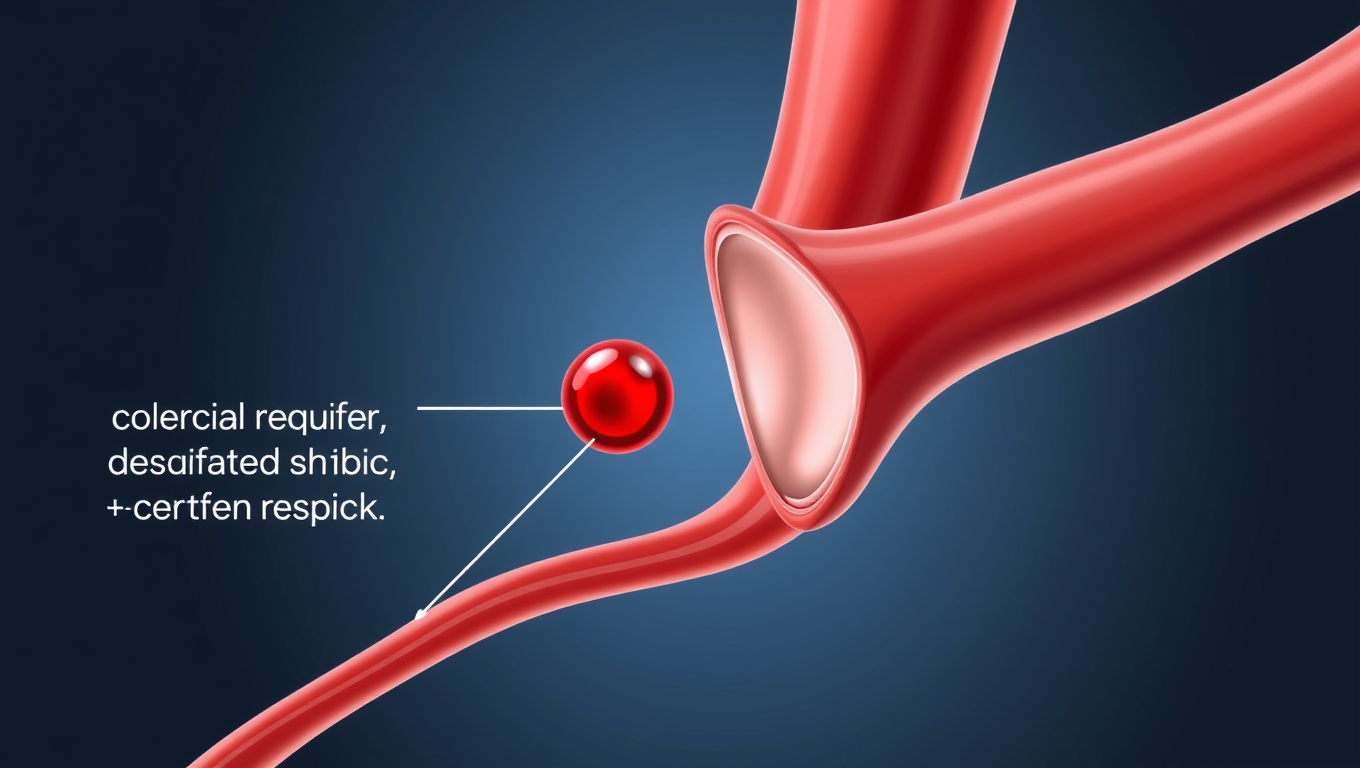
Cervical artery dissection is a tear in an artery in the neck that provides blood flow to the brain. Such a tear can result in blood clots that cause stroke. A new study has found almost a five-fold increase in the number of U.S. hospitalizations for cervical artery dissection over a 15-year period.
Avatar photo

Published

on

The study, published on April 2, 2025, in Neurology®, has revealed a shocking five-fold increase in hospitalizations for cervical artery dissection (CAD) over a 15-year period. This alarming trend emphasizes the need for immediate attention from healthcare professionals, researchers, and the public at large to address this critical cause of stroke.

Cervical artery dissection is a condition where a tear occurs in an artery in the neck that supplies blood to the brain, leading to potentially life-threatening strokes. The study found that the average age of those hospitalized for CAD was 51 years old, with just over half experiencing a stroke at the same time as the dissection.

The researchers analyzed data from 125,102 individuals who were hospitalized for CAD and compared it to U.S. Census data to determine the annual rate of dissections. The results showed an average annual increase of 10% in CAD hospitalizations between 2005 and 2019, with a staggering rise from 11 cases per one million people in 2005 to 46 cases per one million people in 2019.

Furthermore, the study found significant variations in the rate of increase among different racial groups, with Hispanic participants experiencing an average annual increase of 16%, Black participants at 13%, Asian participants at 12%, and white participants at 8%. The researchers also noted a higher average annual increase among individuals aged 65 and older, which stood at 12% compared to 8% for those under 65.

The study’s lead author, Shadi Yaghi, MD, emphasized the importance of detecting CAD right away, as strokes that are not fatal can lead to long-term disability, poor mental health, and reduced quality of life. Dr. Yaghi also highlighted the need for prevention strategies and new treatments to reduce the risk of stroke associated with CAD.

The study’s limitations include the fact that hospital admission data may not account for undiagnosed or untreated cases, which could lead to an even higher incidence of CAD.

In conclusion, the findings of this study serve as a stark reminder of the importance of addressing cervical artery dissection and its association with stroke. The public, healthcare professionals, and researchers must work together to develop effective prevention strategies and treatments to reduce the risk of stroke and improve outcomes for those affected by this condition.

Birth Defects

Unconsciousness by Design: How Anesthetics Shift Brainwave Phase to Induce Slumber

A new study finds that an easily measurable brain wave shift of phase may be a universal marker of unconsciousness under general anesthesia.

Avatar photo

Published

on

The rewritten article aims to make the complex scientific concepts more accessible to a general audience while maintaining the core ideas and findings of the original study.

Unconsciousness by Design: How Anesthetics Shift Brainwave Phase to Induce Slumber

Scientists have long been fascinated by the mysterious world of unconsciousness, trying to understand what happens in our brains when we fall asleep or are anesthetized. A new study has shed light on this phenomenon, revealing a common thread among different anesthetics: they all induce unconsciousness by shifting brainwave phase.

Ketamine and dexmedetomidine, two distinct anesthetics with different molecular mechanisms, were used in the study to demonstrate how these drugs achieve the same result – inducing unconsciousness. By analyzing brain wave activity, researchers found that both anesthetics push around brain waves, causing them to fall out of phase.

In a conscious state, local groups of neurons in the brain’s cortex can share information to produce cognitive functions such as attention, perception, and reasoning. However, when brain waves become misaligned, these local communications break down, leading to unconsciousness.

The study, led by graduate student Alexandra Bardon, discovered that the way anesthetics shift brainwave phase is a potential signature of unconsciousness that can be measured. This finding has significant implications for anesthesiology care, as it could provide a common new measure for anesthesiologists to ensure patients remain unconscious during surgery.

“If you look at the way phase is shifted in our recordings, you can barely tell which drug it was,” said Earl K. Miller, senior author of the study and Picower Professor. “That’s valuable for medical practice.”

The researchers also found that distance played a crucial role in determining the change in phase alignment. Even across short distances, low-frequency waves moved out of alignment, with a 180-degree shift observed between arrays in the upper and lower regions within a hemisphere.

This study raises many opportunities for follow-up research, including exploring how other anesthetics affect brainwave phase and investigating the role of traveling waves in the phenomenon. Furthermore, understanding the difference between anesthesia-induced unconsciousness and sleep could lead to new insights into the mechanisms that generate consciousness.

In conclusion, this study provides a fascinating glimpse into the world of unconsciousness, revealing a common thread among different anesthetics. By continuing to explore the intricacies of brainwave phase alignment, scientists may uncover more secrets about the mysteries of the human brain.

Continue Reading

Accident and Trauma

Lowered Tackle Height Reduces Head Contacts in Women’s Rugby, Study Finds

Lowering the legal tackle height in women’s rugby is proving effective in reducing head contacts between players, a new study suggests. Changes to the tackle height law in women’s community rugby in Scotland is linked to reductions in head-to-head and head-to shoulder contacts, the study found. The researchers used video analysis to study the impact of the lowered tackle height law which World Rugby, the sport’s governing body, introduced for community rugby in an attempt to improve safety for players.

Avatar photo

Published

on

The lowered legal tackle height in women’s community rugby has shown to be effective in reducing head contacts between players, according to a world-first study published in BMJ Open Sport and Exercise Medicine. The research, conducted by researchers at the University of Edinburgh in collaboration with Scottish Rugby and World Rugby, analyzed video footage from 34 Scottish community women’s rugby matches played before and after the introduction of the lowered tackle height law.

The study found that the reduced tackle height led to a significant decrease in head-to-head and head-to-shoulder contacts between players. Specifically, the research revealed:

* A 21% reduction in upright tackles
* A 34% increase in tacklers entering the tackle bent at the waist, which is considered the recommended technique to reduce contact with high-risk areas of the head and shoulders
* A 64% reduction in tacklers making initial contact with the ball carrier’s head and neck
* A 17% reduction in the rate of head-to-head contacts for the tackler
* A 35% reduction in head-to-shoulder contacts for the tackler

The study also found a 19% reduction in contacts above the sternum, known as the “red zone,” between the tackler and the ball carrier. This is considered an area of high risk for concussion.

While the study did not find a significant change in the rate of concussions and injuries when comparing the pre-trial and trial seasons, researchers note that the number of reported injuries overall was very low and may have impacted these findings.

The lead author of the study, Hannah Walton from the University of Edinburgh’s Moray House School of Education and Sport, emphasized the importance of continued collection of robust tackle and injury data to further understand the effect of the law change on player behavior and safety.

This research provides valuable insights into the impact of lowering the tackle height in women’s community rugby and can inform future injury prevention initiatives. The study is part of an international project led by World Rugby to assess the effects of lowering the tackle height in 11 countries, including Australia, England, France, Ireland, Italy, Japan, New Zealand, Scotland, South Africa, and Wales.

Continue Reading

Biochemistry

A Breakthrough in Brain Research: The Iontronic Pipette Revolutionizes Neurological Studies

Researchers have developed a new type of pipette that can deliver ions to individual neurons without affecting the sensitive extracellular milieu. Controlling the concentration of different ions can provide important insights into how individual brain cells are affected, and how cells work together. The pipette could also be used for treatments.

Avatar photo

Published

on

The development of an iontronic pipette at Linköping University has opened up new avenues for neurological research. This innovative tool allows researchers to deliver ions directly to individual neurons without affecting the surrounding extracellular milieu. By controlling the concentration of various ions, scientists can gain valuable insights into how brain cells respond to different stimuli and interact with each other.

The human brain consists of approximately 85-100 billion neurons, supported by a similar number of glial cells that provide essential functions such as nutrition, oxygenation, and healing. The extracellular milieu, a fluid-filled space between the cells, plays a crucial role in maintaining cell function. Changes in ion concentration within this environment can activate or inhibit neuronal activity, making it essential to study how local changes affect individual brain cells.

Previous attempts to manipulate the extracellular environment involved pumping liquid into the area, disrupting the delicate biochemical balance and making it difficult to determine whether the substances themselves or the changed pressure were responsible for the observed effects. To overcome this challenge, researchers at the Laboratory of Organic Electronics developed an iontronic micropipette measuring only 2 micrometers in diameter.

This tiny pipette can deliver ions such as potassium and sodium directly into the extracellular milieu, allowing scientists to study how individual neurons respond to these changes. Glial cell activity is also monitored, providing a more comprehensive understanding of brain function.

Theresia Arbring Sjöström, an assistant professor at LOE, highlighted that glial cells are critical components of the brain’s chemical environment and can be precisely activated using this technology. In experiments conducted on mouse hippocampus tissue slices, it was observed that neurons responded dynamically to changes in ion concentration only after glial cell activity had saturated.

This research has significant implications for neurological disease treatment. The iontronic pipette could potentially be used to develop extremely precise treatments for conditions such as epilepsy, where brain function can be disrupted by localized imbalances in ion concentrations.

Researchers are now continuing their studies on chemical signaling in healthy and diseased brain tissue using the iontronic pipette. They also aim to adapt this technology to deliver medical drugs directly to affected areas of the brain, paving the way for more targeted treatments for neurological disorders.

Continue Reading

Trending