
While we try to keep things accurate, this content is part of an ongoing experiment and may not always be reliable.
Please double-check important details — we’re not responsible for how the information is used.
Health & Medicine
AI Sniffs Out Parkinson’s with 94% Accuracy using Earwax Samples
Imagine diagnosing Parkinson s disease not with pricey scans or subjective checklists, but with a simple ear swab. Scientists in China have developed a promising early screening method that detects Parkinson s from subtle changes in the scent of ear wax yes, really. By analyzing specific volatile compounds in ear wax and feeding that data into an AI-powered olfactory system, they achieved 94% accuracy in identifying who had the disease. If expanded successfully, this low-cost, non-invasive technique could transform early detection and treatment of this debilitating neurological disorder.


Birth Control
Scientists Uncover Groundbreaking Treatment for Resistant High Blood Pressure
A breakthrough pill, baxdrostat, has shown remarkable success in lowering dangerously high blood pressure in patients resistant to standard treatments. In a large international trial, it cut systolic pressure by nearly 10 mmHg, enough to significantly reduce risks of heart attack, stroke, and kidney disease. The drug works by blocking excess aldosterone, a hormone that drives uncontrolled hypertension.

Gastrointestinal Problems
“Cells’ Hidden Shortcut for Healing May Fuel Cancer”
Scientists have uncovered a surprising new healing mechanism in injured cells called cathartocytosis, in which cells “vomit” out their internal machinery to revert more quickly to a stem cell-like state. While this messy shortcut helps tissues regenerate faster, it also leaves behind debris that can fuel inflammation and even cancer.

Arthritis
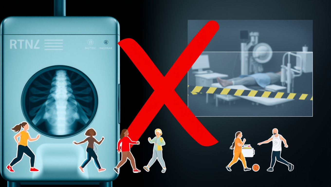
The Alarming Impact of Routine X-Rays on Arthritis Patients’ Decisions
Knee osteoarthritis is a major cause of pain and disability, but routine X-rays often do more harm than good. New research shows that being shown an X-ray can increase anxiety, make people fear exercise, and lead them to believe surgery is the only option, even when less invasive treatments could help. By focusing on clinical diagnosis instead, patients may avoid unnecessary scans, reduce health costs, and make better choices about their care.

A New Horizon for Vision: How Gold Nanoparticles May Restore People’s Sight

Retiring Abroad Can Be Lonely Business

Revolutionizing Quantum Communication: Direct Connections Between Multiple Processors
Harnessing Water Waves: A Breakthrough in Controlling Floating Objects

“Unveiling Hidden Patterns: A New Twist on Interference Phenomena”

Household Electricity Three Times More Expensive Than Upcoming ‘Eco-Friendly’ Aviation E-Fuels, Study Reveals
Reducing Falls Among Elderly Women with Polypharmacy through Exercise Intervention
-
 Detectors12 months ago
A New Horizon for Vision: How Gold Nanoparticles May Restore People’s Sight
-
Earth & Climate1 year ago
Retiring Abroad Can Be Lonely Business
-
Cancer1 year ago
Revolutionizing Quantum Communication: Direct Connections Between Multiple Processors
-
Albert Einstein1 year ago
Harnessing Water Waves: A Breakthrough in Controlling Floating Objects
-
Chemistry1 year ago
“Unveiling Hidden Patterns: A New Twist on Interference Phenomena”
-
Earth & Climate1 year ago
Household Electricity Three Times More Expensive Than Upcoming ‘Eco-Friendly’ Aviation E-Fuels, Study Reveals
-
Diseases and Conditions1 year ago
Reducing Falls Among Elderly Women with Polypharmacy through Exercise Intervention
-
Agriculture and Food1 year ago
“A Sustainable Solution: Researchers Create Hybrid Cheese with 25% Pea Protein”


