Connect with us
We’re experimenting with AI-generated content to help deliver information faster and more efficiently.
While we try to keep things accurate, this content is part of an ongoing experiment and may not always be reliable.
Please double-check important details — we’re not responsible for how the information is used.

Biochemistry

Bringing Clarity to Cancer Genomes with SAVANA: A Machine Learning Algorithm for Long-Read Sequencing

SAVANA uses a machine learning algorithm to identify cancer-specific structural variations and copy number aberrations in long-read DNA sequencing data. The complex structure of cancer genomes means that standard analysis tools give false-positive results, leading to erroneous clinical interpretations of tumour biology. SAVANA significantly reduces such errors. SAVANA offers rapid and reliable genomic analysis to better analyse clinical samples, thereby informing cancer diagnosis and therapeutic interventions.
Avatar photo

Published

on

SAVANA is a groundbreaking algorithm that uses machine learning to accurately identify structural variants and copy number aberrations in cancer genomes. This innovative tool has been developed to overcome the limitations of existing analysis tools, which often fall short when analyzing long-read sequencing data. The complex structure of cancer genomes means that standard analysis tools can lead to false-positive results and unreliable interpretations of the data.

Researchers at EMBL’s European Bioinformatics Institute (EMBL-EBI) and the R&D laboratory of Genomics England have developed SAVANA in collaboration with clinical partners at University College London (UCL), the Royal National Orthopaedic Hospital (RNOH), Instituto de Medicina Molecular João Lobo Antunes, and Boston Children’s Hospital. The algorithm was tested across 99 human tumour samples and has shown remarkable accuracy in distinguishing between true cancer-related genomic alterations and sequencing artefacts.

“SAVANA changes the game,” said Isidro Cortes-Ciriano, Group Leader at EMBL-EBI. “By training the algorithm directly on long-read sequencing data from cancer samples, we created a new method that can tell the difference between true cancer-related genomic alterations and sequencing artefacts, thereby enabling us to elucidate the mutational processes underlying cancer using long-read sequencing with unprecedented resolution.”

The team’s focus was clear: create a tool sophisticated enough to characterise complex cancer genomes but practical enough for clinical use. SAVANA can accurately distinguish somatic structural variants, copy number aberrations, tumour purity, and ploidy – all key to understanding tumour biology and guiding clinical treatment decisions.

Its rapid analysis and robust error correction make SAVANA well suited for clinical use. The method was recently applied to study osteosarcoma, a rare and aggressive bone cancer that mostly affects young people, where it helped researchers uncover new genomic rearrangements, providing novel insights into how osteosarcoma evolves and progresses.

“The capability to accurately detect structural variants is transformative for clinical diagnostics,” said Adrienne Flanagan, Professor at UCL, Consultant Histopathologist at RNOH. “SAVANA could help us confidently identify genomic alterations relevant for diagnosis and prognosis. Ultimately, this means we would be better placed to deliver personalised treatments for cancer patients.”

The UK is investing significantly in genomic sequencing technologies as part of the NHS Genomic Medicine Service. This initiative aims to improve diagnostic accuracy and support personalised cancer treatments. However, investments in clinical genomics will only achieve their intended impact if genomic data are interpreted accurately.

“Using SAVANA will ensure clinicians receive accurate and reliable genomic data, enabling them to confidently integrate advanced genomic sequencing methods such as long-read sequencing into routine patient care,” said Greg Elgar, Director of Sequencing R&D at Genomics England.

SAVANA is being deployed as part of nationwide initiatives, such as the UK Stratified Medicine Paediatrics project funded by Cancer Research UK and Children With Cancer UK, and co-led by Cortes-Ciriano. This project aims to develop more efficacious and less toxic treatments for childhood cancers using advanced sequencing technologies to better understand tumour biology and monitor disease recurrence.

Additionally, SAVANA is being used in Societal, Ancestry, Molecular and Biological Analyses of Inequalities (SAMBAI), a Cancer Grand Challenges funded project aimed at addressing cancer disparities in recent African heritage populations.

Ancient Civilizations

Unlocking the Secrets of Ancient Human Remains: A New Method for Accessing Proteins in Soft Tissues

A new method could soon unlock the vast repository of biological information held in the proteins of ancient soft tissues. The findings could open up a new era for palaeobiological discovery.

Avatar photo

Published

on

By

The article you provided is a fascinating study on a groundbreaking method for extracting and identifying proteins from ancient human soft tissues. Here’s a rewritten version, maintaining the core ideas but improving clarity, structure, and style:

Unlocking the Secrets of Ancient Human Remains: A New Method for Accessing Proteins in Soft Tissues

A team of researchers at the University of Oxford has developed a revolutionary method that could soon unlock the vast repository of biological information held in the proteins of ancient human soft tissues. This discovery, published in PLOS ONE, opens up a new era for palaeobiological discovery and promises to vastly expand our understanding of ancient diet, disease, environment, and evolutionary relationships.

Up until now, studies on ancient proteins have been confined largely to mineralized tissues such as bones and teeth. However, the internal organs – which are a far richer source of biological information – have remained inaccessible due to the lack of an established protocol for their analysis. This new method changes that.

A key hurdle was finding an effective way to disrupt cell membranes to liberate proteins. The team discovered that urea successfully broke open cells and released proteins within. After extraction, the proteins were then separated using liquid chromatography and identified using mass spectrometry. By coupling this step with high-field asymmetric-waveform ion mobility spectrometry (which separates ions based on how they move in an electric field), the researchers found that they could increase the number of proteins identified by up to 40%.

This technique makes it possible to recover proteins from samples that are hard to analyze, including degraded or very complex mixtures. The team was able to identify over 1,200 ancient proteins from just 2.5 mg of sample – a feat that has never been achieved before.

Using the combined method, the researchers identified a diverse array of proteins that govern healthy brain function, reflecting the molecular complexity of the human nervous system. They also identified potential biomarkers for neurological diseases such as Alzheimer’s and multiple sclerosis. This new technique opens a window on human history we haven’t looked through before.

The vast majority of human diseases – including psychiatric illness and mental health disorders – leave no marks on the bone, making them essentially invisible in the archaeological record. This discovery promises to transform our understanding of ancient human health and disease.

Senior author Professor Roman Fischer, Centre for Medicines Discovery at the University of Oxford, added: “By enabling the retrieval of protein biomarkers from ancient soft tissues, this workflow allows us to investigate pathology beyond the skeleton, transforming our ability to understand the health of past populations.”

This method has already attracted interest for its applicability to a wide range of archaeological materials and environments – from mummified remains to bog bodies, and from antibodies to peptide hormones. As Dr Christiana Scheib, Department of Zoology at the University of Cambridge, noted: “Ancient soft tissues are so rarely preserved, yet could hold such powerful information regarding evolutionary history.”

Continue Reading

Air Quality

“Visualizing the Rotten Egg: Scientists Use Cryo-EM to Reveal the 3D Structure of Sulfite Reductase”

Most people have witnessed — or rather smelled — when a protein enzyme called sulfite reductase works its magic. This enzyme catalyzes the chemical reduction of sulfite to hydrogen sulfide. Hydrogen sulfide is the rotten egg smell that can occur when organic matter decays and is frequently associated with sewage treatment facilities and landfills. But scientists have not been able to capture a visual image of the enzyme’s structure until now, thus limiting their full understanding of how it works.

Avatar photo

Published

on

By

The ability to visualize complex chemical reactions has long been a holy grail for scientists. For one particularly pungent protein enzyme called sulfite reductase, this dream has finally become a reality, thanks to the work of Florida State University Professor Elizabeth Stroupe and her former doctoral student Behrouz Ghazi Esfahani.

Sulfite reductase is an enzyme that catalyzes the reduction of sulfite to hydrogen sulfide, which is infamous for its “rotten egg” smell. This reaction occurs in various natural environments, from fruit and vegetable decomposition to sewage treatment facilities and landfills. However, despite its importance, scientists had been unable to capture a clear visual image of the enzyme’s structure – until now.

Using an advanced technique called cryo-electron microscopy (cryo-EM), Stroupe and Ghazi Esfahani were able to visualize the 3D structure of sulfite reductase in unprecedented detail. Cryo-EM allows scientists to capture images of chemical reactions as they occur, providing the necessary data to reconstruct the complex molecular structures.

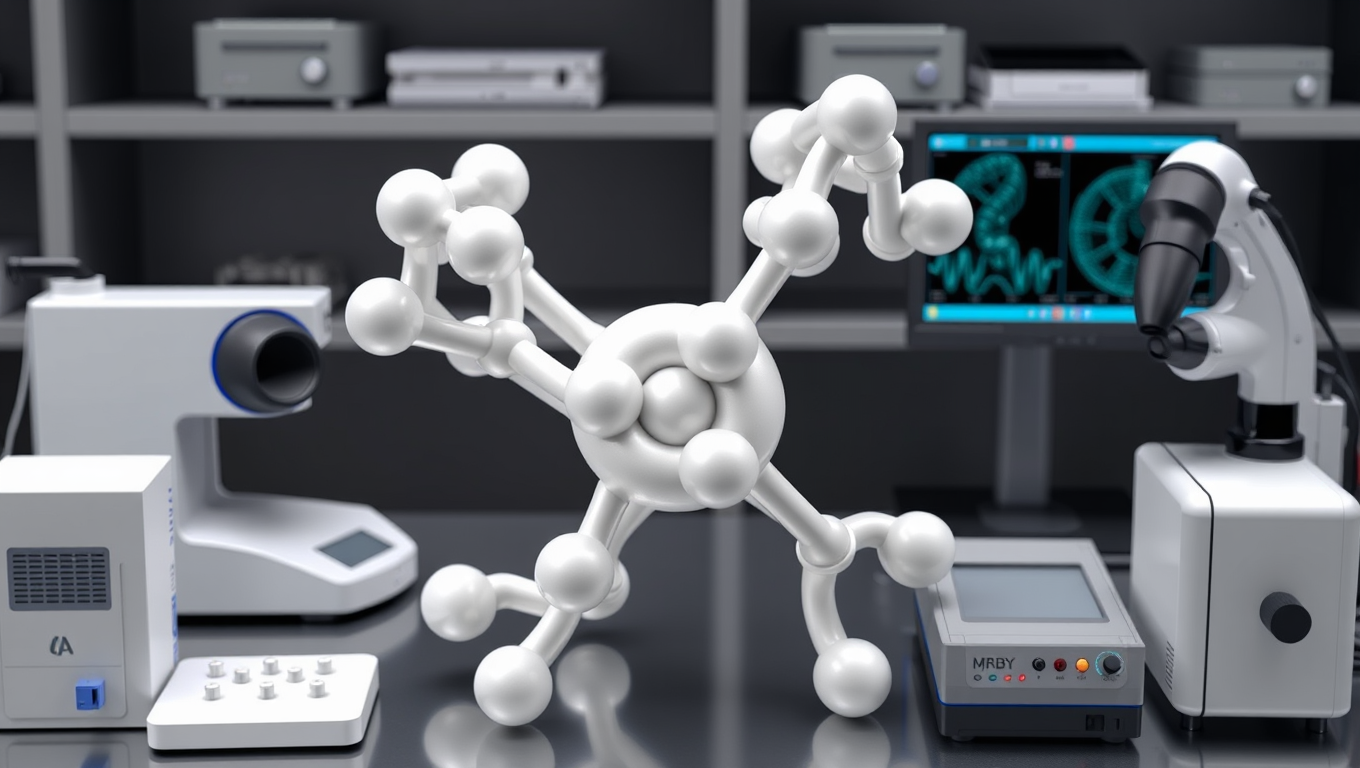
The resulting image is a striking representation of the protein’s intricate arrangement of atoms and electron transfer mechanisms. Stroupe describes it as an “octopus with four yo-yos” due to its flexibility and dynamic nature.

This breakthrough has significant implications for scientists, particularly in understanding how to control or manipulate chemical reactions – a process crucial for drug manufacturers and industry. As Ghazi Esfahani notes, the research also has environmental implications, such as understanding how bacteria use sulfur as an energy source.

While this achievement marks a major step forward in understanding sulfite reductase, there are still unanswered questions about its function as part of larger protein assemblies and how similar enzymes work in other organisms – like the pathogen that causes tuberculosis. Stroupe’s lab is continuing to explore these mysteries, shedding more light on the intricate chemistry of sulfur metabolism.

In conclusion, the ability to visualize complex chemical reactions has finally been achieved for sulfite reductase, thanks to the innovative use of cryo-EM by Stroupe and Ghazi Esfahani. This breakthrough opens doors to new understanding and manipulation of chemical reactions – with far-reaching implications for science, industry, and the environment.

Continue Reading

Batteries

The Cellular Gatekeepers: How Membranes May Have Shaped Life on Earth

A team of researchers studied the properties of membranes to understand how these cellular structures influenced the chemistry of life on Earth as it began.

Avatar photo

Published

on

The origin of life on Earth remains one of science’s most enduring mysteries. Researchers have long sought to understand how the first cells emerged from the primordial soup, and what properties these early membranes may have had is a crucial piece of this puzzle. By studying the features shared among all life today, scientists can better grasp how life began and evolved into the incredible diversity we see in organisms today.

One essential feature of membranes is their selective permeability – determining which molecules pass through and which are kept out. This has a significant impact on the biological processes that keep cells functioning. Researchers have focused on three types of molecules crucial for all life: sugars, amino acids, and nucleic acids. These molecules are vital because they exhibit chirality – a property where molecules can twist in specific ways, much like our left and right hands.

In biology, chirality is critical for how molecules interact with each other. For example, the sugars in DNA and RNA must all have the same chirality (right-handed) to assemble into the backbone of a strand. However, the reason life chose one chirality over the other has remained a long-standing question.

A recent study proposes that early membranes may have played a key role in selecting the right-handed sugars and left-handed amino acids that are used by all life today. The researchers analyzed what molecules could pass through membranes with properties similar to those of archaea, a major group of microbes. They also designed a membrane that combined properties of both archaeal and bacterial membranes.

The results showed that right-handed DNA and RNA sugars more easily passed through these membranes, while left-handed versions had trouble permeating. There was more variability among amino acids, with some left-handed versions being able to pass through the mixed bacterial and archaeal membrane. This included alanine, one of the first amino acids thought to be used by life.

These findings demonstrate how differences in membranes can strongly affect which molecules are able to pass through. Since the membranes studied are only approximations of what the first life on Earth may have been encased in, there may be other unknown properties of the earliest membranes that influenced our most essential molecules.

The authors conclude, “All known life uses a specific stereochemistry: left-handed amino acids and right-handed DNA. Understanding how this evolved is a long-standing mystery key for understanding the origin of life. Our experiments show that a specific type of membrane – the structure that encloses cells – acts as a sieve that selects for the stereochemistry life uses.”

Continue Reading

Trending