
While we try to keep things accurate, this content is part of an ongoing experiment and may not always be reliable.
Please double-check important details — we’re not responsible for how the information is used.
Biochemistry
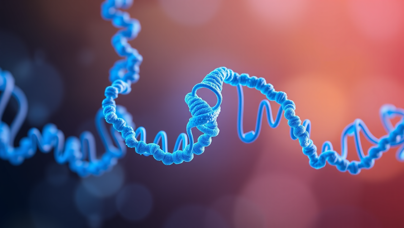
Designing Enzymes from Scratch: A Breakthrough in Chemistry
Researchers have developed a new workflow for designing enzymes from scratch, paving the way toward more efficient, powerful and environmentally benign chemistry. The new method allows designers to combine a variety of desirable properties into new-to-nature catalysts for an array of applications, from drug development to materials design.


Biochemistry
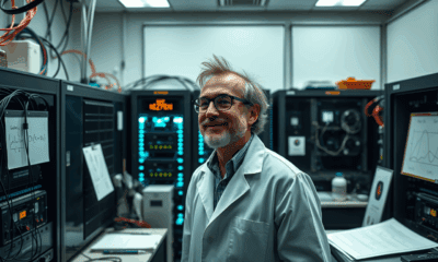
Shape-Shifting Catalysts: Revolutionizing Green Chemistry with a Single Atom
A team in Milan has developed a first-of-its-kind single-atom catalyst that acts like a molecular switch, enabling cleaner, more adaptable chemical reactions. Stable, recyclable, and eco-friendly, it marks a major step toward programmable sustainable chemistry.
Biochemistry
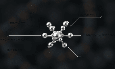
Scientists Finally Tame the Impossible: A Stable 48-Atom Carbon Ring is Achieved
Researchers have synthesized a stable cyclo[48]carbon, a unique 48-carbon ring that can be studied in solution at room temperature, a feat never achieved before.
Biochemistry
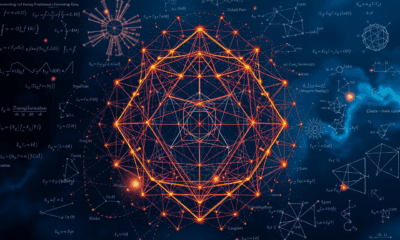
“Revolutionizing Medicine: A 100x Faster Path to Life-Saving Drugs with Metal Carbenes”
Using a clever combo of iron and radical chemistry, scientists have unlocked a safer, faster way to create carbenes molecular powerhouses key to modern medicine and materials. It s 100x more efficient than previous methods.

A New Horizon for Vision: How Gold Nanoparticles May Restore People’s Sight

Retiring Abroad Can Be Lonely Business

Revolutionizing Quantum Communication: Direct Connections Between Multiple Processors
Harnessing Water Waves: A Breakthrough in Controlling Floating Objects

“Unveiling Hidden Patterns: A New Twist on Interference Phenomena”

Household Electricity Three Times More Expensive Than Upcoming ‘Eco-Friendly’ Aviation E-Fuels, Study Reveals

“A Sustainable Solution: Researchers Create Hybrid Cheese with 25% Pea Protein”
-
 Detectors11 months ago
A New Horizon for Vision: How Gold Nanoparticles May Restore People’s Sight
-
Earth & Climate1 year ago
Retiring Abroad Can Be Lonely Business
-
Cancer1 year ago
Revolutionizing Quantum Communication: Direct Connections Between Multiple Processors
-
Albert Einstein1 year ago
Harnessing Water Waves: A Breakthrough in Controlling Floating Objects
-
Chemistry12 months ago
“Unveiling Hidden Patterns: A New Twist on Interference Phenomena”
-
Earth & Climate1 year ago
Household Electricity Three Times More Expensive Than Upcoming ‘Eco-Friendly’ Aviation E-Fuels, Study Reveals
-
Agriculture and Food1 year ago
“A Sustainable Solution: Researchers Create Hybrid Cheese with 25% Pea Protein”
-
Diseases and Conditions1 year ago
Reducing Falls Among Elderly Women with Polypharmacy through Exercise Intervention


