
While we try to keep things accurate, this content is part of an ongoing experiment and may not always be reliable.
Please double-check important details — we’re not responsible for how the information is used.
Diabetes
Digital PCR Revolutionizes Chronic Myeloid Leukemia Treatment: A Breakthrough for Patients in Remission
Researchers have found that the clinical application of BCR::ABL1 digital PCR can reliably quantify stable deep molecular remission of chronic myeloid leukemia (CML), which will help to determine for which patients chronic drug treatment could potentially be discontinued. This transcript that is unique for CML is more sensitive and accurate than the current standard, real-time quantitative PCR (RT-qPCR), for detecting ultralow levels of residual leukemic disease.


Allergy
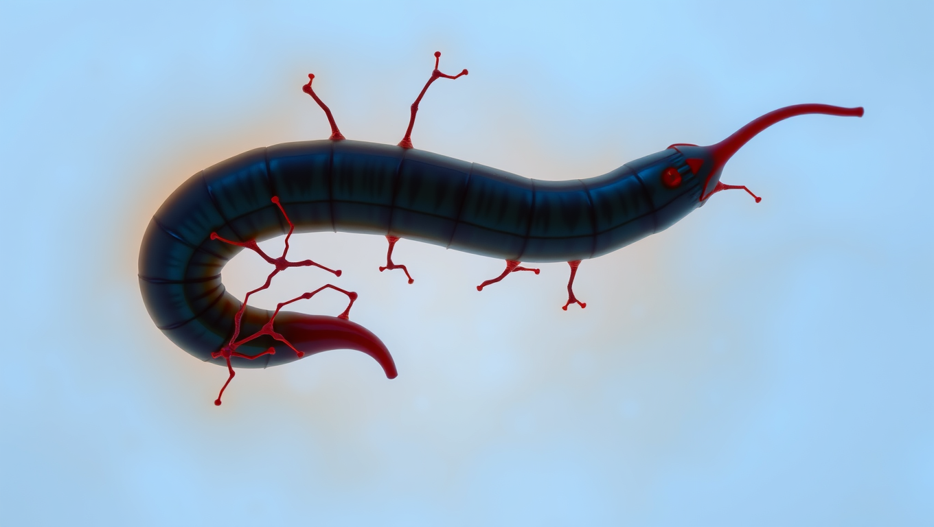
“The Silent Invader: How a Parasitic Worm Evades Detection and What it Can Teach Us About Pain Relief”
Scientists have discovered a parasite that can sneak into your skin without you feeling a thing. The worm, Schistosoma mansoni, has evolved a way to switch off the body’s pain and itch signals, letting it invade undetected. By blocking certain nerve pathways, it avoids triggering the immune system’s alarms. This stealth tactic not only helps the worm survive, but could inspire new kinds of pain treatments and even preventative creams to protect people from infection.
Colon Cancer
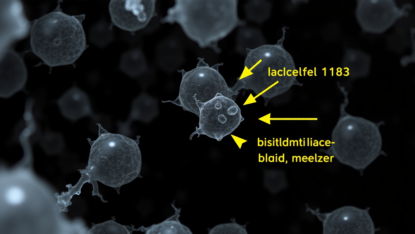
Scientists Discover a Tiny Molecule That Could Revolutionize Weight Loss Treatment
Researchers at the Salk Institute have used CRISPR to uncover hidden microproteins that control fat cell growth and lipid storage, identifying one confirmed target, Adipocyte-smORF-1183. This breakthrough could lead to more effective obesity treatments, surpassing the limitations of current drugs like GLP-1.
Chronic Illness
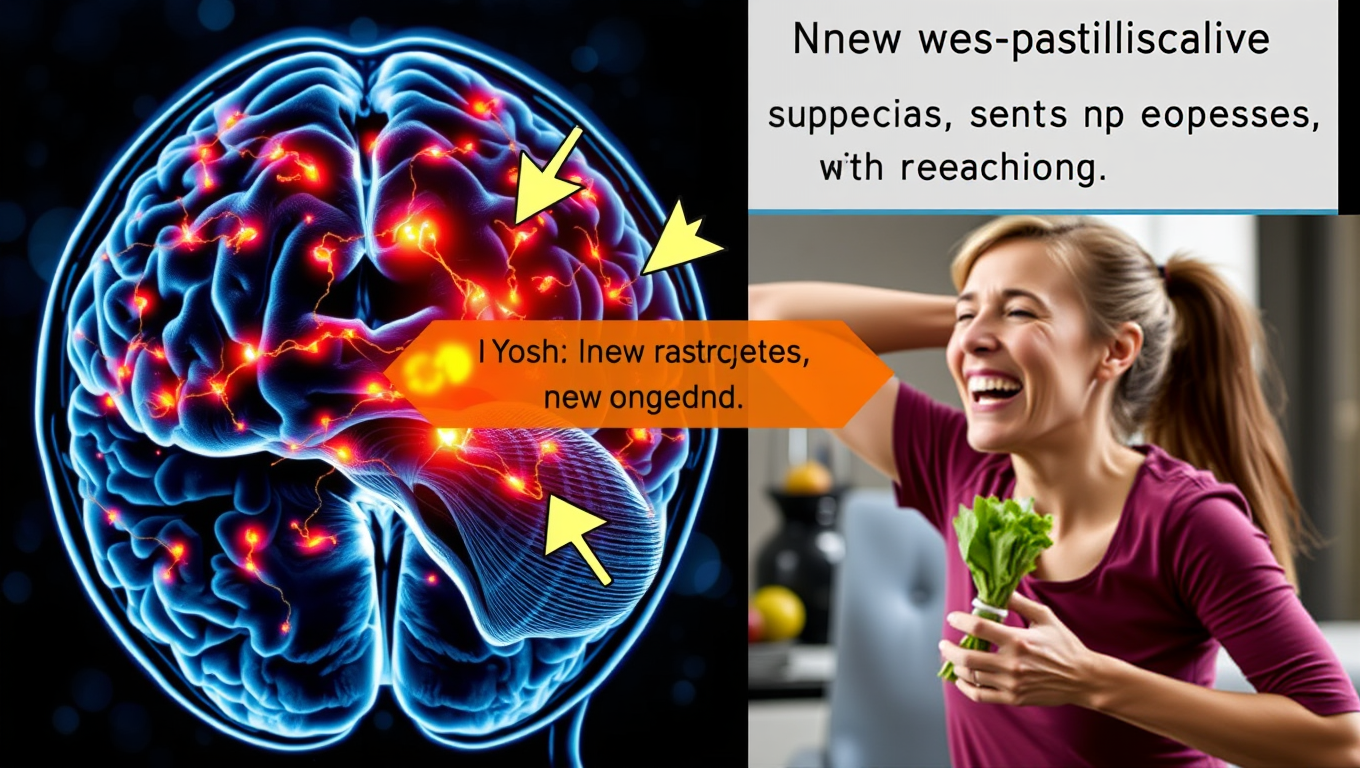
Scientists Uncover Hidden Brain Shortcut for Weight Loss without Nausea
Scientists have uncovered a way to promote weight loss and improve blood sugar control without the unpleasant side effects of current GLP-1 drugs. By shifting focus from neurons to brain support cells that produce appetite-suppressing molecules, they developed a modified compound, TDN, that worked in animal tests without causing nausea or vomiting.

A New Horizon for Vision: How Gold Nanoparticles May Restore People’s Sight

Retiring Abroad Can Be Lonely Business

Revolutionizing Quantum Communication: Direct Connections Between Multiple Processors
Harnessing Water Waves: A Breakthrough in Controlling Floating Objects

“Unveiling Hidden Patterns: A New Twist on Interference Phenomena”

Household Electricity Three Times More Expensive Than Upcoming ‘Eco-Friendly’ Aviation E-Fuels, Study Reveals
Reducing Falls Among Elderly Women with Polypharmacy through Exercise Intervention
-
 Detectors12 months ago
A New Horizon for Vision: How Gold Nanoparticles May Restore People’s Sight
-
Earth & Climate1 year ago
Retiring Abroad Can Be Lonely Business
-
Cancer1 year ago
Revolutionizing Quantum Communication: Direct Connections Between Multiple Processors
-
Albert Einstein1 year ago
Harnessing Water Waves: A Breakthrough in Controlling Floating Objects
-
Chemistry1 year ago
“Unveiling Hidden Patterns: A New Twist on Interference Phenomena”
-
Earth & Climate1 year ago
Household Electricity Three Times More Expensive Than Upcoming ‘Eco-Friendly’ Aviation E-Fuels, Study Reveals
-
Diseases and Conditions1 year ago
Reducing Falls Among Elderly Women with Polypharmacy through Exercise Intervention
-
Agriculture and Food1 year ago
“A Sustainable Solution: Researchers Create Hybrid Cheese with 25% Pea Protein”


