
While we try to keep things accurate, this content is part of an ongoing experiment and may not always be reliable.
Please double-check important details — we’re not responsible for how the information is used.
Developmental Biology
“Evolution of Defense: How Cells Adapt to Malicious Jumping Genes”
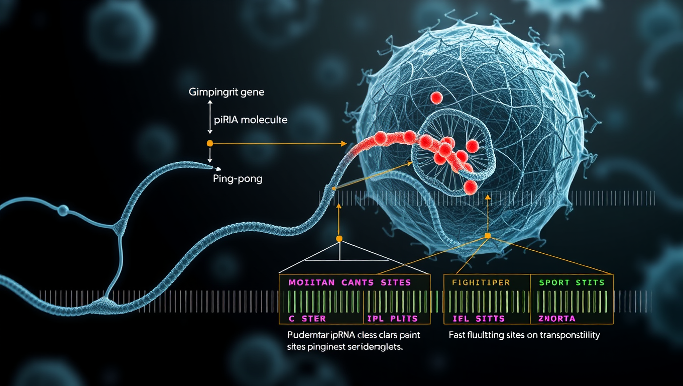
Adverse genetic mutations can cause harm and are due to various circumstances. ‘Jumping genes’ are one cause of mutations, but cells try and combat them with a specialized RNA called piRNA. Researchers have identified how the sites responsible for piRNA production evolve effective behaviors against jumping genes. This research could lead to downstream diagnostic or therapeutic applications.


Behavioral Science
“Rewired for Romance: Scientists Give Gift-Giving Behavior to Singing Fruit Flies”
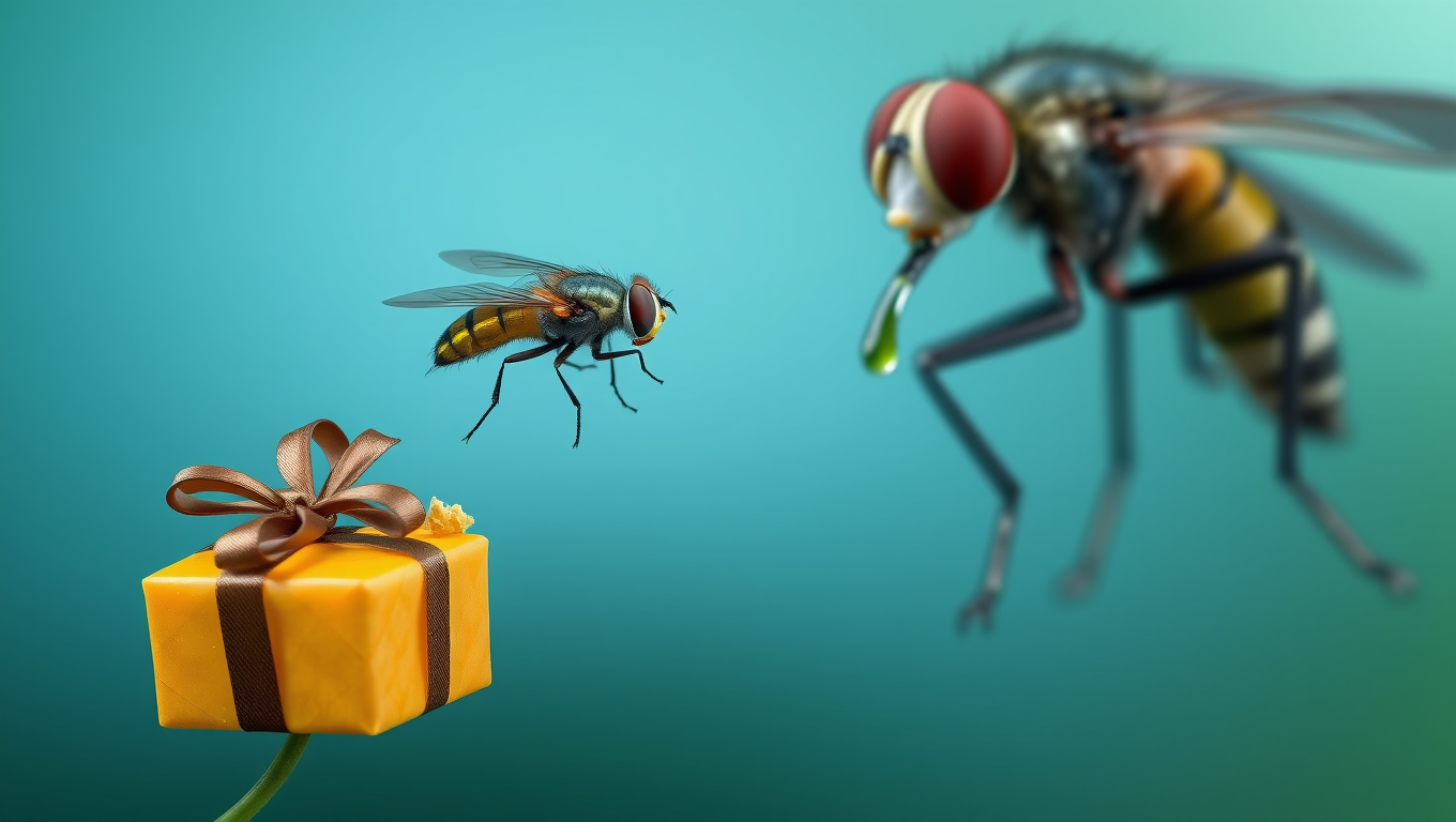
By flipping a single genetic switch, researchers made one fruit fly species adopt the gift-giving courtship of another, showing how tiny brain rewiring can drive evolutionary change.
Agriculture and Food
Breaking New Ground: Scientists Develop Groundbreaking Chromosome Editing Technology
A group of Chinese scientists has created powerful new tools that allow them to edit large chunks of DNA with incredible accuracy—and without leaving any trace. Using a mix of advanced protein design, AI, and clever genetic tweaks, they’ve overcome major limitations in older gene editing methods. These tools can flip, remove, or insert massive pieces of genetic code in both plants and animals. To prove it works, they engineered rice that’s resistant to herbicides by flipping a huge section of its DNA—something that was nearly impossible before.
Biochemistry Research
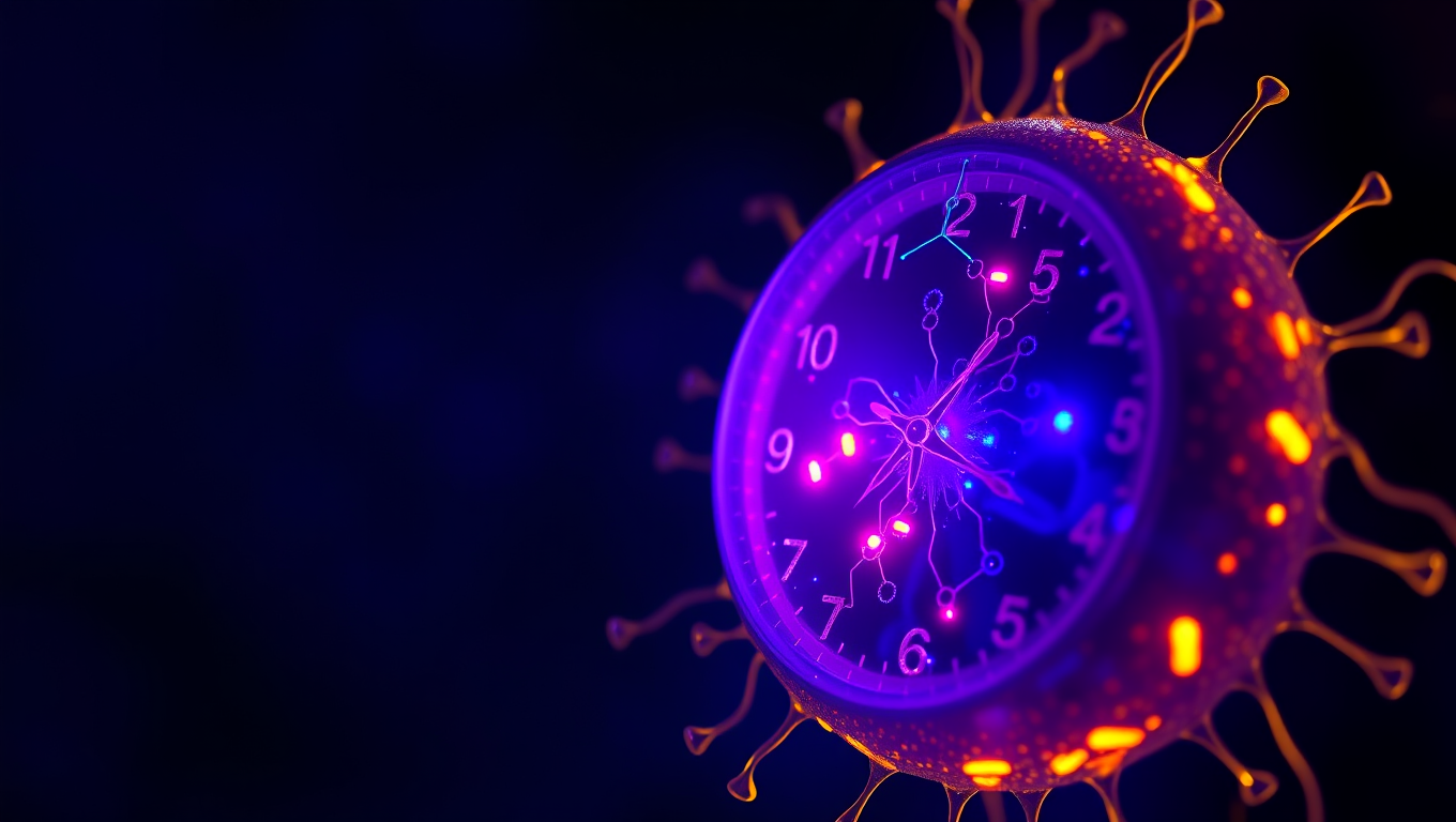
“Unlocking Timekeeping Secrets: Scientists Reveal How Artificial Cells Can Accurately Keep Rhythm”
Scientists at UC Merced have engineered artificial cells that can keep perfect time—mimicking the 24-hour biological clocks found in living organisms. By reconstructing circadian machinery inside tiny vesicles, the researchers showed that even simplified synthetic systems can glow with a daily rhythm—if they have enough of the right proteins.

A New Horizon for Vision: How Gold Nanoparticles May Restore People’s Sight

Retiring Abroad Can Be Lonely Business

Revolutionizing Quantum Communication: Direct Connections Between Multiple Processors
Harnessing Water Waves: A Breakthrough in Controlling Floating Objects

“Unveiling Hidden Patterns: A New Twist on Interference Phenomena”

Household Electricity Three Times More Expensive Than Upcoming ‘Eco-Friendly’ Aviation E-Fuels, Study Reveals
Reducing Falls Among Elderly Women with Polypharmacy through Exercise Intervention
-
 Detectors11 months ago
A New Horizon for Vision: How Gold Nanoparticles May Restore People’s Sight
-
Earth & Climate1 year ago
Retiring Abroad Can Be Lonely Business
-
Cancer12 months ago
Revolutionizing Quantum Communication: Direct Connections Between Multiple Processors
-
Albert Einstein1 year ago
Harnessing Water Waves: A Breakthrough in Controlling Floating Objects
-
Chemistry12 months ago
“Unveiling Hidden Patterns: A New Twist on Interference Phenomena”
-
Earth & Climate12 months ago
Household Electricity Three Times More Expensive Than Upcoming ‘Eco-Friendly’ Aviation E-Fuels, Study Reveals
-
Diseases and Conditions1 year ago
Reducing Falls Among Elderly Women with Polypharmacy through Exercise Intervention
-
Agriculture and Food1 year ago
“A Sustainable Solution: Researchers Create Hybrid Cheese with 25% Pea Protein”


