While we try to keep things accurate, this content is part of an ongoing experiment and may not always be reliable.
Please double-check important details — we’re not responsible for how the information is used.
Biotechnology and Bioengineering
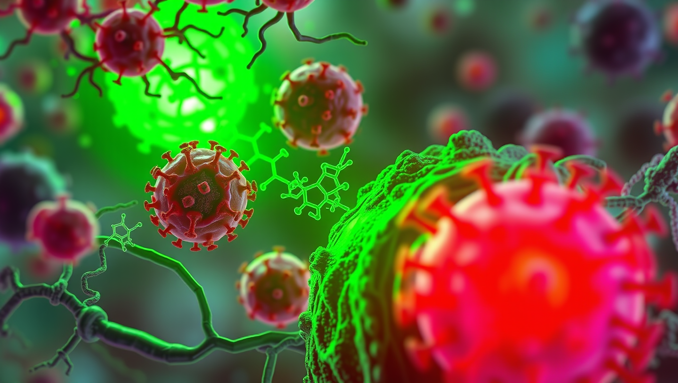
Gut Microbes Unleash Cancer-Fighting Bile Acids with Anti-Androgen Properties
Bacteria naturally present in the human intestine (known as the gut microbiota) can transform cholesterol-derived bile acids into powerful metabolites that strengthen anti-cancer immunity by blocking androgen signaling, according to a preclinical study.


Behavioral Science
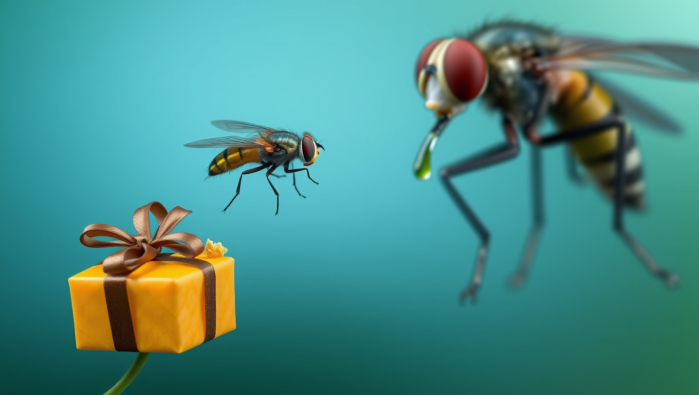
“Rewired for Romance: Scientists Give Gift-Giving Behavior to Singing Fruit Flies”
By flipping a single genetic switch, researchers made one fruit fly species adopt the gift-giving courtship of another, showing how tiny brain rewiring can drive evolutionary change.
Biotechnology and Bioengineering
A Trojan Horse Approach: Bacteria-Delivered Viruses Show Promise in Cancer Treatment
Scientists have engineered a groundbreaking cancer treatment that uses bacteria to smuggle viruses directly into tumors, bypassing the immune system and delivering a powerful one-two punch against cancer cells. The bacteria act like Trojan horses, carrying viral payloads to cancer’s core, where the virus can spread and destroy malignant cells. Built-in safety features ensure the virus can’t multiply outside the tumor, offering a promising pathway for safe, targeted therapy.

Artificial Intelligence
Accelerating Evolution: The Power of T7-ORACLE in Protein Engineering
Researchers at Scripps have created T7-ORACLE, a powerful new tool that speeds up evolution, allowing scientists to design and improve proteins thousands of times faster than nature. Using engineered bacteria and a modified viral replication system, this method can create new protein versions in days instead of months. In tests, it quickly produced enzymes that could survive extreme doses of antibiotics, showing how it could help develop better medicines, cancer treatments, and other breakthroughs far more quickly than ever before.


A New Horizon for Vision: How Gold Nanoparticles May Restore People’s Sight

Revolutionizing Quantum Communication: Direct Connections Between Multiple Processors

Retiring Abroad Can Be Lonely Business
Harnessing Water Waves: A Breakthrough in Controlling Floating Objects

“Unveiling Hidden Patterns: A New Twist on Interference Phenomena”

Household Electricity Three Times More Expensive Than Upcoming ‘Eco-Friendly’ Aviation E-Fuels, Study Reveals
Reducing Falls Among Elderly Women with Polypharmacy through Exercise Intervention
-
 Detectors1 year ago
A New Horizon for Vision: How Gold Nanoparticles May Restore People’s Sight
-
Cancer1 year ago
Revolutionizing Quantum Communication: Direct Connections Between Multiple Processors
-
Earth & Climate1 year ago
Retiring Abroad Can Be Lonely Business
-
Albert Einstein1 year ago
Harnessing Water Waves: A Breakthrough in Controlling Floating Objects
-
Chemistry1 year ago
“Unveiling Hidden Patterns: A New Twist on Interference Phenomena”
-
Earth & Climate1 year ago
Household Electricity Three Times More Expensive Than Upcoming ‘Eco-Friendly’ Aviation E-Fuels, Study Reveals
-
Diseases and Conditions1 year ago
Reducing Falls Among Elderly Women with Polypharmacy through Exercise Intervention
-
Earth & Climate1 year ago
Predicting Damage from Local Earthquakes in Mexico City