Connect with us
We’re experimenting with AI-generated content to help deliver information faster and more efficiently.
While we try to keep things accurate, this content is part of an ongoing experiment and may not always be reliable.
Please double-check important details — we’re not responsible for how the information is used.

Bone and Spine

Harnessing Ocean Energy: Top Locations Revealed for Clean Power Generation Worldwide

Until now, a global evaluation of ocean current energy with actual data was lacking. Using 30 years of NOAA’s Global Drifter Program data, a study shows that ocean currents off Florida’s East Coast and South Africa have exceptionally high-power densities, ideal for electricity generation. With densities over 2,500 watts per square meter, these regions are 2.5 times more energy-dense than ‘excellent’ wind resources. Shallow waters further enhance the potential for ocean current turbines, unlike areas like Japan and South America, which have lower densities at similar depths.

Avatar photo

Published

on

The quest for sustainable energy sources has led researchers to explore the vast potential of oceans. Covering over 70% of our planet’s surface, oceans offer an enormous opportunity for clean energy production from renewable resources like ocean currents and waves. However, marine renewable energy development is still in its early stages compared to wind and solar power. To overcome this challenge, scientists have been working on identifying the most feasible locations for ocean current energy projects.

A groundbreaking study by researchers at Florida Atlantic University has shed light on the best places globally for harnessing clean energy from ocean currents. Using over 30 years of data from NOAA’s Global Drifter Program (GDP), they analyzed more than 43 million data points to provide the most comprehensive assessment of ocean current energy to date.

The study revealed that areas with high power densities are ideal for generating electricity from ocean currents. Specifically, waters off Florida’s East coast and South Africa consistently showed power densities above 2,500 watts per square meter, which is more energy-dense than an “excellent” wind energy resource.

“We found that about 75% of the total high-power density areas have energy levels between 500 and 1,000 watts per square meter. This suggests there’s a lot of potential for harvesting energy from ocean currents,” said Mahsan Sadoughipour, Ph.D., first author and graduate research assistant in the College of Engineering and Computer Science.

Other key findings include:

* Regions with high power densities are generally found off the Southeast coast of the U.S. (Florida to North Carolina), as well as along the Eastern and Southeastern coasts of Africa.
* Lower power densities were seen in areas like Japan, Northern South America (Brazil and French Guiana), and the Eastern coast of Australia.
* The accuracy of power density estimates was highly reliable in regions like North America and Japan but less reliable in areas with limited data or highly variable water conditions.

The study also highlighted the importance of considering factors such as seasonal variations, environmental conditions, and measurement methods when estimating ocean current energy potential.

This groundbreaking research has significant implications for the global transition to renewable energy. By identifying optimal locations for ocean current energy projects, we can better integrate this clean source into our broader energy landscape.

As Gabriel Alsenas, director of FAU’s Southeast National Marine Renewable Energy Center, noted, “This research further solidifies Southeast Florida as one of the premier locations for harnessing the power of ocean currents.”

Alternative Medicine

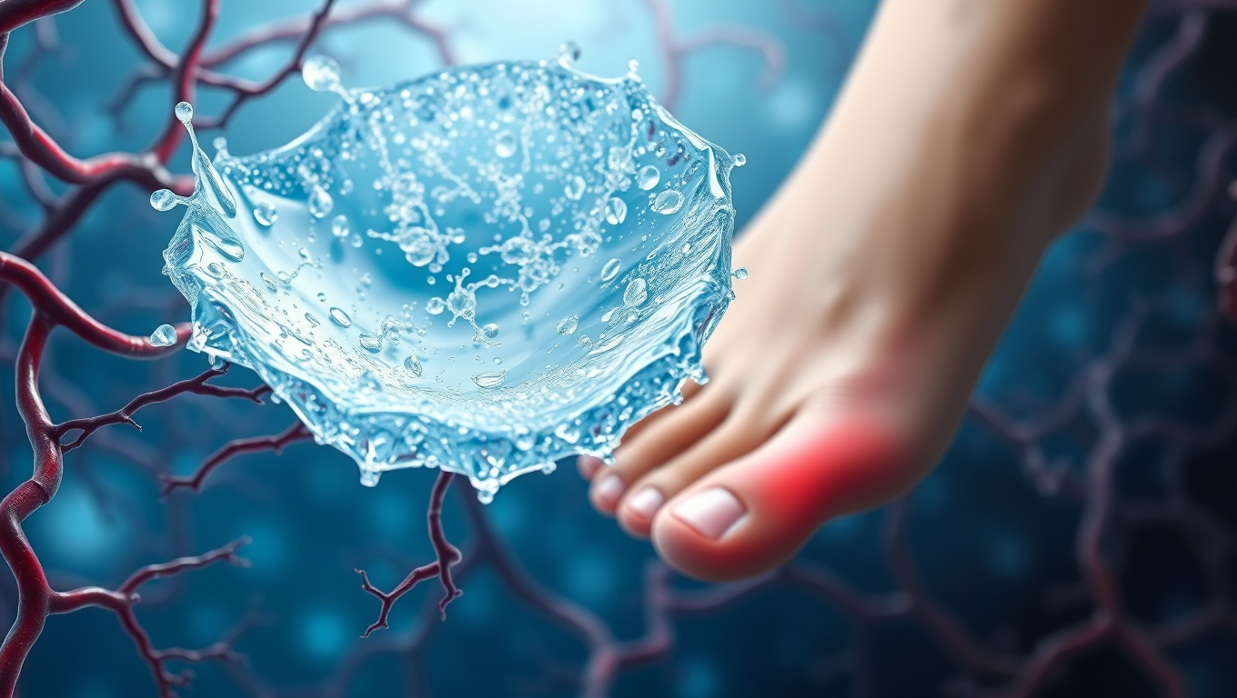
Breaking Barriers in Diabetic Wound Healing: A Revolutionary “Smart” Gel Accelerates Blood Flow and Restores Tissue Repair

A new gel-based treatment could change the way diabetic wounds heal. By combining tiny healing messengers called vesicles with a special hydrogel, scientists have created a dressing that restores blood flow and helps wounds close much faster. In tests, the treatment healed diabetic wounds far quicker than normal, while also encouraging the growth of new blood vessels. Researchers believe this innovation could one day help millions of people with slow-healing wounds caused by diabetes and possibly other conditions.

Avatar photo

Published

on

Breaking Barriers in Diabetic Wound Healing: A Revolutionary “Smart” Gel Accelerates Blood Flow and Restores Tissue Repair

Chronic diabetic wounds, particularly diabetic foot ulcers, pose a significant burden for patients due to impaired blood vessel growth and subsequent tissue repair issues. A groundbreaking study has unveiled a novel approach by combining small extracellular vesicles (sEVs) loaded with miR-221-3p and a GelMA hydrogel to target thrombospondin-1 (TSP-1), a protein that suppresses angiogenesis. This innovative bioactive wound dressing not only accelerates healing but also promotes blood vessel formation, offering a promising new approach to treating one of the most challenging complications of diabetes.

The study explores a new method to stimulate angiogenesis and speed up the healing process by targeting TSP-1 with miR-221OE-sEVs encapsulated in GelMA. This engineered hydrogel has shown significant enhancement in wound healing and blood vessel formation in diabetic mice, offering hope for more effective treatments in the future.

Researchers discovered that high glucose conditions commonly found in diabetic wounds lead to increased levels of TSP-1 in endothelial cells, impairing their ability to proliferate and migrate – key processes for angiogenesis. By utilizing miR-221-3p, a microRNA that targets and downregulates TSP-1 expression, they restored endothelial cell function. The engineered miR-221OE-sEVs were encapsulated within a GelMA hydrogel, ensuring a controlled release at the wound site.

In animal trials, this composite dressing dramatically accelerated wound healing, with a notable increase in vascularization and a 90% wound closure rate within just 12 days, compared to slower healing in control groups. This breakthrough has significant implications for diabetic wound care, offering patients more efficient and lasting wound healing solutions.

As further research and clinical trials progress, the promise of combining miRNA-based therapies with biocompatible hydrogels could become a cornerstone in regenerative medicine, opening up possibilities beyond diabetic foot ulcers. The technology could be adapted for use in treating other chronic wounds, such as those caused by vascular diseases, or even in regenerating tissues like bone and cartilage.

Continue Reading

Bone and Spine

The Quiet Side Effect of Semaglutide: How Losing Muscle Can Affect Weight Loss Success

Semaglutide, a popular anti-obesity drug, may come with a hidden cost: significant muscle loss, especially in women and older adults. A small study found that up to 40% of weight loss from semaglutide comes from lean body mass. Alarmingly, those who consumed less protein saw even more muscle loss—potentially undermining improvements in blood sugar control.

Avatar photo

Published

on

In recent years, the anti-obesity medication semaglutide has gained popularity for its ability to melt fat effectively. However, a small study presented at ENDO 2025, the Endocrine Society’s annual meeting in San Francisco, highlights a concerning side effect of this medication – the potential quiet stripping away of muscle strength, particularly among women and older adults.

Losing muscle mass, also known as lean mass, is a common issue associated with weight loss in adults with obesity. This can have negative consequences on metabolism and bone health. Muscle plays an essential role in controlling blood sugar levels after meals and keeping bones strong. Approximately 40% of the weight lost from taking semaglutide comes from lean mass, including muscle.

The researchers conducted a three-month study involving 40 adults with obesity, divided into two groups: those prescribed semaglutide and those following a diet and lifestyle program called Healthy Habits for Life (HHL). The results showed that participants who took semaglutide lost more weight than those in the HHL group. However, the percentage of weight loss attributed to lean mass was similar between the two groups.

Further analysis revealed that being older, female, or consuming less protein was linked to greater muscle loss among participants taking semaglutide. Moreover, losing more muscle was associated with less improvement in blood sugar control (HbA1c levels).

Study lead researcher Melanie Haines emphasized the importance of preserving muscle during weight loss with semaglutide to reduce insulin resistance and prevent frailty in people with obesity. She also highlighted that higher protein intake may help mitigate muscle loss in these patients.

In conclusion, while semaglutide is an effective medication for weight loss, it’s essential to be aware of its potential side effects, particularly among vulnerable populations like women and older adults. By understanding the importance of preserving muscle mass during weight loss and incorporating higher protein intake into their diet, individuals can maximize the benefits of this medication and achieve a healthier, more sustainable weight loss outcome.

Continue Reading

Bone and Spine

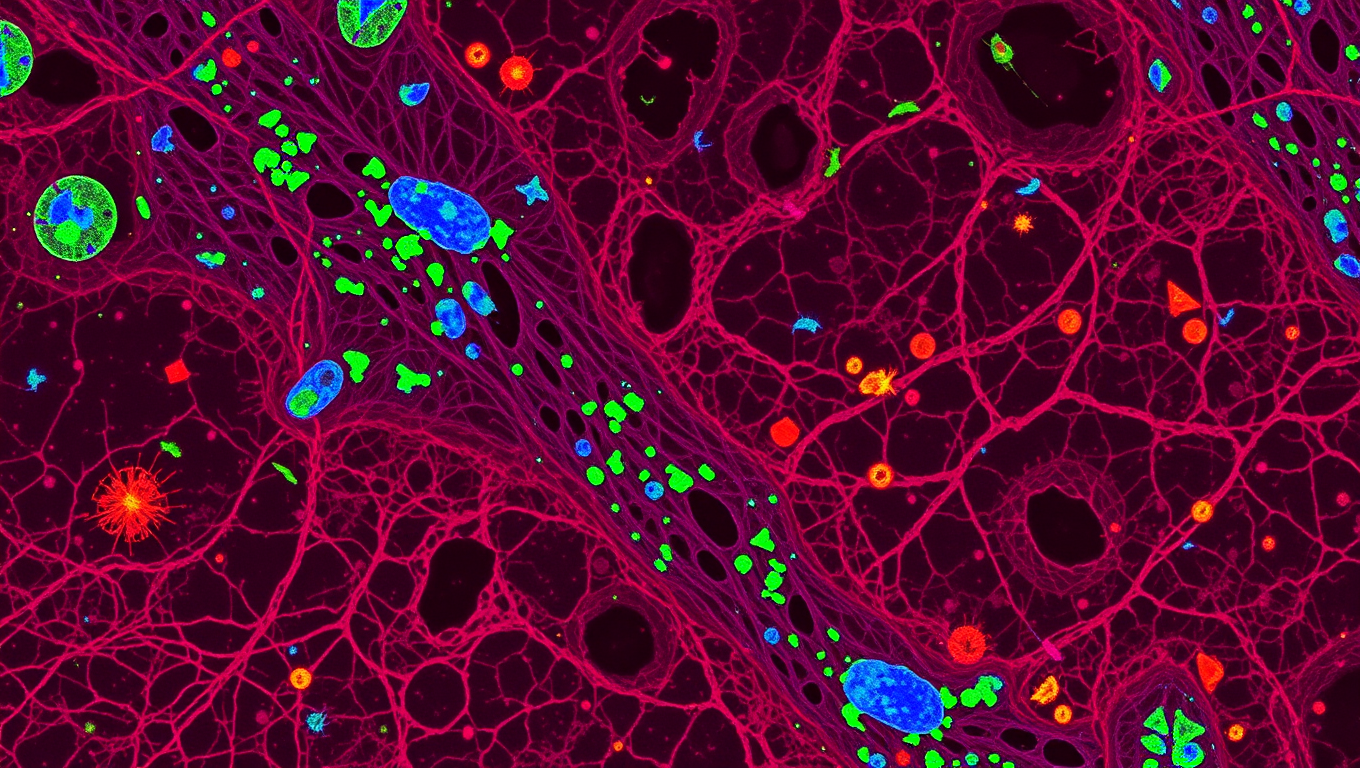
Unlocking New Insights into Bone Marrow: Scientists Develop Revolutionary Imaging Technique

A new bone marrow imaging technique could change treatment for cancer, autoimmune disease and musculoskeletal disorders.

Avatar photo

Published

on

The Indiana University School of Medicine has made a groundbreaking discovery in the field of medical research. A team of scientists has developed a revolutionary new imaging technique that allows for the visualization of bone marrow in mouse models with unprecedented clarity and precision. This breakthrough could have far-reaching implications for the development of new treatments and therapies for conditions affecting bone marrow, such as cancers, autoimmune diseases, and musculoskeletal disorders.

The new method utilizes the multiplex imaging tool Phenocycler 2.0, which enables researchers to visualize a record number of cellular markers within intact bone marrow tissue from mice. This is a significant advancement over traditional methods like flow cytometry, which requires disrupting complex tissues to study and quantify cell populations, and standard fluorescence imaging, which is limited to detecting only three cellular markers at a time.

“We are thrilled to have made this breakthrough,” said Sonali Karnik, PhD, assistant research professor of orthopedic surgery at the IU School of Medicine and co-lead author of the study. “Bone marrow is a complex tissue that plays an essential role in blood and immune cell formation, and housing valuable stem cells. Our new imaging approach offers a unique tool for a variety of research applications.”
The IU Cooperative Center of Excellence in Hematology team successfully applied the Phenocycler 2.0 tool to mouse bone marrow, expanding its capabilities beyond organs like the spleen and kidney. This technique has the potential to revolutionize our understanding of diseases affecting bone marrow and enable researchers to develop more effective treatments.

“The use of mouse models is widespread in studying human diseases,” said Reuben Kapur, PhD, a co-senior author on the study and director of the IU School of Medicine’s Herman B Wells Center for Pediatric Research. “This technique offers a promising new method for investigating conditions like autoimmune diseases, leukemia, and other disorders involving bone marrow.”
The IU Innovation and Commercialization Office has filed a provisional patent for the new imaging methodology, and the team is now working to expand the marker panel to include additional features such as bone, nerves, muscle, and more immune and signaling cell types.

This research was supported by funding from the National Institutes of Health, and its findings were recently published in Leukemia. The development of this revolutionary imaging technique has the potential to transform our understanding of bone marrow and lead to breakthroughs in treating conditions affecting it.

Continue Reading

Trending