Connect with us
We’re experimenting with AI-generated content to help deliver information faster and more efficiently.
While we try to keep things accurate, this content is part of an ongoing experiment and may not always be reliable.
Please double-check important details — we’re not responsible for how the information is used.

Alzheimer's Research

Microplastics in Medical Infusion Bags: A Hidden Health Risk

Microplastics have been found almost everywhere that scientists have looked for them. Now these bits of plastic — from 1 to 62 micrometers long — have been found in the filtered solutions used for medical intravenous (IV) infusions. The researchers estimate that thousands of plastic particles could be delivered directly to a person’s bloodstream from a single 8.4-ounce (250-milliliter) bag of infusion fluid.

Avatar photo

Published

on

The article reveals that microplastics, tiny pieces of plastic measuring 1-62 micrometers in length, have been found in filtered solutions used for medical intravenous (IV) infusions. Researchers estimate that thousands of these particles could be delivered directly into a person’s bloodstream from a single IV bag. This hidden health risk is especially concerning given the widespread use of IV infusions in clinical settings.

The study, published in the Environment & Health journal, analyzed two different brands of 8.4-ounce IV saline solution bags and found that both contained microplastic particles made from polypropylene, the same material as the bags themselves. This suggests that the bags shed microplastics into the solutions, which could potentially enter the bloodstream and cause negative health effects.

The researchers estimate that each bag of infusion fluid could deliver about 7,500 microplastics directly into the bloodstream, with this figure rising to around 25,000 particles for dehydration treatment or 52,500 for abdominal surgery. This is a concerning finding given the potential long-term effects on human health.

To mitigate this risk, the researchers recommend keeping IV infusion bags away from ultraviolet light and heat, as these conditions can cause microplastic shedding. They also suggest using micrometer-level filtration systems to remove particles during infusion.

While there are no clinical studies to date that have assessed the health risks of microplastics exposure, the researchers believe their findings will help inform policies and measures to mitigate potential threats to human health. The study’s authors acknowledge funding from the National Natural Science Foundation of China.

Alzheimer's

Uncovering the Hidden Culprits Behind Alzheimer’s Disease

A surprising new study has uncovered over 200 misfolded proteins in the brains of aging rats with cognitive decline, beyond the infamous amyloid and tau plaques long blamed for Alzheimer’s. These shape-shifting proteins don’t clump into visible plaques, making them harder to detect but potentially just as harmful. Scientists believe these “stealth” molecules could evade the brain’s cleanup systems and quietly impair memory and brain function. The discovery opens a new frontier in understanding dementia and could lead to entirely new targets for treatment and prevention.

Avatar photo

Published

on

Uncovering the Hidden Culprits Behind Alzheimer’s Disease

For decades, researchers have been trying to understand the root causes of Alzheimer’s disease. While amyloids, such as A-beta and tau proteins, have long been the focus of attention, a new study suggests that these sticky brain plaques may not be the only culprits behind cognitive decline.

Researchers at Johns Hopkins University have made a groundbreaking discovery, identifying over 200 types of misfolded proteins in rats that could contribute to age-related cognitive decline. This finding has significant implications for Alzheimer’s research and opens up new avenues for potential therapeutic targets and treatments.

“We’re seeing hundreds of proteins misfolding in ways that don’t clump together in an amyloid and yet still seem to impact how the brain functions,” said Stephen Fried, an assistant professor of chemistry and protein scientist. “Our research is showing that amyloids are just the tip of the iceberg.”

To reach this conclusion, Fried and his team studied 17 two-year-old rats with varying levels of cognitive impairment. They measured over 2,500 types of protein in the hippocampus, a part of the brain associated with spatial learning and memory. The researchers were able to determine which proteins misfolded for all the rats and are associated with aging in general versus which proteins specifically misfold in cognitively impaired rats.

More than 200 proteins were found to be misfolded in the cognitively impaired rats but maintained their shapes in the cognitively healthy rats. This suggests that some of these misfolded proteins may contribute to cognitive decline, according to the researchers.

Misfolded proteins are unable to carry out tasks necessary for a cell to function properly, so cells have a natural surveillance system that identifies and destroys these misbehaving proteins. However, it appears that some misfolded proteins can escape this surveillance system and still cause problems.

The next step for Fried’s team is to use high-resolution microscopes to get a more detailed picture of what the misfolded proteins look like at the molecular level.

“A lot of us have experienced a loved one or a relative who has become less capable of doing those everyday tasks that require cognitive abilities,” Fried said. “Understanding what’s physically going on in the brain could lead to better treatments and preventive measures.”

This research has significant implications for Alzheimer’s disease, as it suggests that there may be multiple targets for treatment beyond amyloids alone. By understanding the molecular differences between healthy and cognitively impaired brains, researchers can develop more effective treatments and potentially prevent cognitive decline in the first place.

Continue Reading

Alzheimer's

Uncovering the Hidden Defenses Against Alzheimer’s Disease: A Breakthrough Study on Brain Resilience

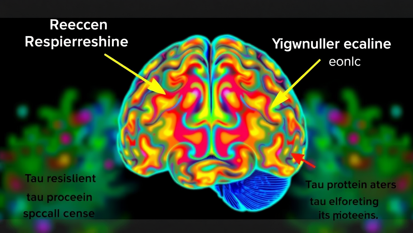
Scientists at UCSF combined advanced brain-network modeling, genetics, and imaging to reveal how tau protein travels through neural highways and how certain genes either accelerate its toxic journey or shield brain regions from damage. Their extended Network Diffusion Model pinpoints four gene categories that govern vulnerability or resilience, reshaping our view of Alzheimer’s progression and spotlighting fresh therapeutic targets.

Avatar photo

Published

on

Alzheimer’s disease is a complex condition that affects different parts of the brain in various ways. One key factor in the progression of the disease is the misbehavior of tau proteins, which can lead to toxic clumps forming inside neurons and impairing their function. Researchers have long sought to understand why some areas of the brain are more resilient to Alzheimer’s than others, a phenomenon known as selective vulnerability or resilience.

A recent study by researchers at the University of California, San Francisco (UCSF) has made significant strides in this area by combining advanced mathematical modeling with brain imaging and genetics. The study, published in Brain, identified multiple distinct pathways through which risk genes confer vulnerability or resilience to Alzheimer’s disease.

The researchers developed a model called the extended Network Diffusion Model (eNDM), which predicted where tau protein would spread next based on real-world brain connection data from healthy individuals. By applying this model to brain scans of 196 people at various stages of Alzheimer’s, they were able to identify areas that were resistant or vulnerable to the disease.

The study revealed four distinct types of genes: those that boost tau spread along the brain’s wiring (Network-Aligned Vulnerability), those that promote tau buildup in ways unrelated to connectivity (Network-Independent Vulnerability), those that help protect regions that are otherwise tau hotspots (Network-Aligned Resilience), and those that offer protection outside of the network’s usual path (Network-Independent Resilience).

These findings have significant implications for understanding Alzheimer’s disease and developing potential intervention targets. The study’s lead author, Ashish Raj, PhD, noted that their research offers a “hopeful map forward” in understanding and eventually stopping Alzheimer’s disease.

The researchers also highlighted the importance of considering the different biological functions of genes that respond independently of the network versus those that respond in concert with it. This nuanced approach could lead to more effective strategies for identifying potential intervention targets and developing treatments for Alzheimer’s disease.

Continue Reading

Alternative Medicine

A Pain-Free Patch Revolutionizes Cancer Detection with Nanoneedles

A new nanotechnology breakthrough may soon eliminate the need for painful biopsies. Scientists have developed a patch filled with nanoneedles thinner than a human hair that can painlessly extract molecular data from tissues without removing or damaging them. This enables real-time disease monitoring, particularly for conditions like brain cancer and Alzheimer s, and could radically change how doctors diagnose and track disease. The patch works quickly, integrates with common medical tools, and provides results using AI, opening doors to personalized medicine and better surgical decisions.

Avatar photo

Published

on

The article has been rewritten to improve clarity, structure, and style, making it understandable to the general public:

A groundbreaking patch, containing tens of millions of microscopic nanoneedles, could soon replace traditional biopsies. This innovative technology offers a painless and less invasive alternative for millions of patients worldwide who undergo biopsies each year to detect and monitor diseases like cancer and Alzheimer’s.

Biopsies are among the most common diagnostic procedures worldwide, performed millions of times every year. However, they can be invasive, cause pain and complications, and deter patients from seeking early diagnosis or follow-up tests. Traditional biopsies also remove small pieces of tissue, limiting how often and how comprehensively doctors can analyze diseased organs like the brain.

Now, scientists at King’s College London have developed a nanoneedle patch that painlessly collects molecular information from tissues without removing or damaging them. This breakthrough could allow healthcare teams to monitor disease in real-time and perform multiple, repeatable tests from the same area – something impossible with standard biopsies.

The nanoneedles are incredibly thin, measuring 1,000 times thinner than a human hair, and cause no pain or damage. For many patients, this means earlier diagnosis and more regular monitoring, transforming how diseases are tracked and treated.

Dr. Ciro Chiappini, who led the research published in Nature Nanotechnology, said: “We have been working on nanoneedles for twelve years, but this is our most exciting development yet. It opens a world of possibilities for people with brain cancer, Alzheimer’s, and for advancing personalized medicine.”

The patch is covered in tens of millions of nanoneedles that extract molecular “fingerprints” – including lipids, proteins, and mRNAs – from cells without harming the tissue. The tissue imprint is then analyzed using mass spectrometry and artificial intelligence, giving healthcare teams detailed insights into whether a tumor is present, how it’s responding to treatment, and how disease is progressing at the cellular level.

This technology could be used during brain surgery to help surgeons make faster, more precise decisions. For example, by applying the patch to a suspicious area, results could be obtained within 20 minutes and guide real-time decisions about removing cancerous tissue.

Made using the same manufacturing techniques as computer chips, the nanoneedles can be integrated into common medical devices such as bandages, endoscopes, and contact lenses. Dr. Chiappini added: “This could be the beginning of the end for painful biopsies. Our technology opens up new ways to diagnose and monitor disease safely and painlessly – helping doctors and patients make better, faster decisions.”

Continue Reading

Trending