Connect with us
We’re experimenting with AI-generated content to help deliver information faster and more efficiently.
While we try to keep things accurate, this content is part of an ongoing experiment and may not always be reliable.
Please double-check important details — we’re not responsible for how the information is used.

Biotechnology

Mysterious Microorganisms Shape Marble and Limestone with Tiny Tubes

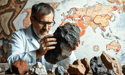
In the desert areas of Namibia, Oman, and Saudi Arabia, research work has revealed unusual structures that are probably due to the activity of an unknown microbiological life form. Unusually small burrows, i.e., tiny tubes that run through the rock in a parallel arrangement from top to bottom, were discovered in marble and limestone of these desert regions.

Avatar photo

Published

on

In the arid regions of Namibia, Oman, and Saudi Arabia, researchers have stumbled upon an enigmatic phenomenon that challenges our understanding of geological processes. Unusually small burrows, or tiny tubes, have been discovered in marble and limestone rocks, which are believed to be the result of microorganisms at work. The discovery was made by Professor Cees Passchier from Johannes Gutenberg University Mainz (JGU), who first encountered this phenomenon during his fieldwork in Namibia.

Passchier’s team has found similar structures in Oman and Saudi Arabia, with the tubes forming bands up to ten meters long. These tiny tunnels are not empty; they are filled with a fine powder of clean calcium carbonate, which is believed to be a remnant of the microorganisms’ activities. The researchers speculate that these microbes may have bored the tunnels to access nutrients present in the calcium carbonate, the main component of marble.

The age of these structures is estimated to be around one or two million years old, with Passchier suggesting that they were formed in a slightly more humid climate than the current desert conditions. However, the microorganisms responsible for creating these tubes remain unknown.

This phenomenon has sparked interest among scientists due to its potential implications on the global carbon cycle. The release of carbon through the biological activity of microorganisms could play a significant role in the Earth’s CO2 balance. As Passchier emphasizes, it is essential that the scientific community becomes aware of this discovery and continues to investigate the mystery surrounding these enigmatic tubes.

In conclusion, the discovery of mysterious microorganisms shaping marble and limestone with tiny tubes offers a fascinating glimpse into the complexities of geological processes. While much remains unknown about these structures and their creators, further research may shed light on the secrets hidden within the Earth’s ancient rocks.

Biochemistry Research

A More Realistic Look at DNA in Action: Researchers Shed Light on the Mechanical Forces Behind Strand Separation

By creating a more true-to-life representation of DNA’s environment, researchers have discovered that strand separation may take more mechanical force than the field previously believed.

Avatar photo

Published

on

The way we study DNA has long been a topic of discussion among researchers. Traditionally, biochemistry labs isolate DNA within a water-based solution that allows scientists to manipulate it without interacting with other molecules. However, this approach can be misleading, as it doesn’t reflect the true environment of a living cell. In fact, the interior of a cell is “super crowded” with molecules, which can significantly impact the behavior of DNA.

Researchers at Northwestern University have taken a more realistic approach to studying DNA by creating an environment that mimics the conditions within a living cell. Led by Professor John Marko, the team used microscopic magnetic tweezers to separate DNA and then carefully attach strands of it to surfaces on one end, and tiny magnetic particles on the other. This allowed them to conduct high-tech imaging and investigate how different types of molecules interact with DNA.

The researchers found that strand separation, a crucial process for initiating replication or making repairs, may require more mechanical force than previously believed. They introduced three types of molecules to the solution holding DNA, mimicking proteins and investigating interactions among glycerol, ethylene glycol, and polyethylene glycol (each approximately the size of one DNA double helix, two or three nanometers).

“We wanted to have a wide variety of molecules where some cause dehydration, destabilizing DNA mechanically, and then others that stabilize DNA,” said Northwestern post-doctoral researcher Parth Desai. “It’s not exactly analogous to things found in cells, but you could imagine that other competing proteins in cells will have a similar effect.”
The team wrote a paper on their findings, which will be published on June 17 in the Biophysical Journal. Marko and Desai hope to run more experiments that incorporate multiple crowding agents and move closer to a true representation of a cell.

“If this affects DNA strand separation, all protein interactions with DNA are also going to be affected,” said Marko. “For example, the tendency for proteins to stick to specific sites on DNA and to control specific processes — this is also going to be altered by crowding.”

Their research has significant implications for understanding fundamental biochemical processes and may lead to new medical advances. The team hopes to study how interactions between enzymes and DNA are impacted by crowding in a living cell, which could have far-reaching consequences for our understanding of cellular biology.

This work was supported by the National Institutes of Health (grant R01-GM105847) and by subcontract to the University of Massachusetts Center for 3D Structure and Physics of the Genome (under NIH grant UM1-HG011536).

Continue Reading

Biotechnology

Breaking Ground: Ultrasound Revolutionizes Hydrogel Manufacturing

Researchers have developed a new way to create hydrogels using ultrasound, eliminating the need for toxic chemical initiators. This breakthrough offers a faster, cleaner and more sustainable approach to hydrogel fabrication, and produces hydrogels that are stronger, more flexible and highly resistant to freezing and dehydration. The new method also promises to facilitate advances in tissue engineering, bioadhesives and 3D bioprinting.

Avatar photo

Published

on

The world of hydrogel manufacturing has just gotten a whole lot greener. Researchers at McGill University, in collaboration with Polytechnique Montréal, have pioneered a groundbreaking method to create hydrogels using ultrasound, eliminating the need for toxic chemical initiators. This innovation promises a faster, cleaner, and more sustainable approach to hydrogel fabrication, producing materials that are stronger, more flexible, and highly resistant to freezing and dehydration.

Hydrogels, composed of polymers that can absorb and retain large amounts of water, have numerous applications in wound dressings, drug delivery, tissue engineering, soft robotics, and more. Traditional hydrogel manufacturing relies on chemical initiators, some of which can be hazardous, particularly in medical applications. These chemicals trigger chemical chain reactions, but the McGill research team has developed an alternative method using ultrasound.

When applied to a liquid precursor, sound waves create microscopic bubbles that collapse with immense energy, triggering gel formation within minutes. This ultrasound-driven technique is dubbed “sonogel.” According to Mechanical Engineering Professor Jianyu Li, who led the research team, the problem they aimed to solve was the reliance on toxic chemical initiators.

“Our method eliminates these substances, making the process safer for the body and better for the environment,” said Li. With sonogel, gel formation occurs in just five minutes, compared to hours or even overnight under UV light. This speed and efficiency have significant implications for biomedical applications.

One of the most exciting possibilities for this technology is in non-invasive medical treatments. Because ultrasound waves can penetrate deep into tissues, this method could enable in-body hydrogel formation without surgery. Imagine injecting a liquid precursor and using ultrasound to solidify it precisely where needed – this could be a game-changer for treating tissue damage and regenerative medicine.

Further refinement of this technique also opens the door to ultrasound-based 3D bioprinting. Instead of relying on light or heat, researchers could use sound waves to precisely “print” hydrogel structures. By leveraging high-intensity focused ultrasound, researchers can shape and build hydrogels with remarkable precision.

According to Jean Provost, one of co-authors of the study and assistant professor of engineering physics at Polytechnique Montréal, this breakthrough has significant potential for safer, greener material production. The sonogel method has the potential to revolutionize biomedical applications and unlock new possibilities for non-invasive medical treatments, making it a truly groundbreaking innovation in the field of hydrogel manufacturing.

Continue Reading

Bacteria

Unlocking Efficiency: Researchers Reveal Secrets of Cell Division with Min Proteins

The Min protein system prevents abnormal cell division in bacteria, but is poorly understood. Researchers have uncovered how engineered e.coli bacteria control protein levels for maximum efficiency.

Avatar photo

Published

on

The Min protein system is a complex process that helps bacteria divide evenly and correctly. For decades, scientists have studied this system, but understanding how it works efficiently has been a challenge. Recently, researchers at the University of California San Diego (UCSD) made a groundbreaking discovery that sheds new light on the efficiency of cell division.

The UCSD team developed a way to control Min protein expression levels independently in E. coli cells. This allowed them to observe how different concentrations of Min proteins affect the oscillations between the poles of the cell. The results were surprising: despite varying concentrations, the oscillations remained stable across a wide range, with E. coli producing just the right amount of Min proteins.

This breakthrough is significant because it shows that the Min protein system can efficiently guide division to the correct location without relying on precise control over protein levels. This finding has far-reaching implications for our understanding of cellular organization and function.

The study was published in Nature Physics, a leading scientific journal, and was funded by the National Institutes of Health (NIH). The research team consisted of experts from both physics and chemistry/biochemistry departments at UCSD, highlighting the importance of interdisciplinary collaboration in advancing our knowledge of cellular biology.

Continue Reading

Trending