While we try to keep things accurate, this content is part of an ongoing experiment and may not always be reliable.
Please double-check important details — we’re not responsible for how the information is used.
Biochemistry
“Reading Cancer’s Molecular Signatures: A Breakthrough in Early Detection”
Cancer diagnoses traditionally require invasive or labor-intensive procedures such as tissue biopsies. Now, research reveals a method that uses pulsed infrared light to identify molecular profiles in blood plasma that could indicate the presence of certain common cancers. In this proof-of-concept study, blood plasma from more than 2,000 people was analyzed to link molecular patterns to lung cancer, extrapolating a potential ‘cancer fingerprint.’


Biochemistry
Shape-Shifting Catalysts: Revolutionizing Green Chemistry with a Single Atom
A team in Milan has developed a first-of-its-kind single-atom catalyst that acts like a molecular switch, enabling cleaner, more adaptable chemical reactions. Stable, recyclable, and eco-friendly, it marks a major step toward programmable sustainable chemistry.
Biochemistry
Scientists Finally Tame the Impossible: A Stable 48-Atom Carbon Ring is Achieved
Researchers have synthesized a stable cyclo[48]carbon, a unique 48-carbon ring that can be studied in solution at room temperature, a feat never achieved before.

Biochemistry
“Revolutionizing Medicine: A 100x Faster Path to Life-Saving Drugs with Metal Carbenes”
Using a clever combo of iron and radical chemistry, scientists have unlocked a safer, faster way to create carbenes molecular powerhouses key to modern medicine and materials. It s 100x more efficient than previous methods.


A New Horizon for Vision: How Gold Nanoparticles May Restore People’s Sight

Revolutionizing Quantum Communication: Direct Connections Between Multiple Processors

Retiring Abroad Can Be Lonely Business

“Unveiling Hidden Patterns: A New Twist on Interference Phenomena”
Harnessing Water Waves: A Breakthrough in Controlling Floating Objects

Household Electricity Three Times More Expensive Than Upcoming ‘Eco-Friendly’ Aviation E-Fuels, Study Reveals
Reducing Falls Among Elderly Women with Polypharmacy through Exercise Intervention
-
 Detectors1 year ago
A New Horizon for Vision: How Gold Nanoparticles May Restore People’s Sight
-
Cancer1 year ago
Revolutionizing Quantum Communication: Direct Connections Between Multiple Processors
-
Earth & Climate1 year ago
Retiring Abroad Can Be Lonely Business
-
Chemistry1 year ago
“Unveiling Hidden Patterns: A New Twist on Interference Phenomena”
-
Albert Einstein1 year ago
Harnessing Water Waves: A Breakthrough in Controlling Floating Objects
-
Earth & Climate1 year ago
Household Electricity Three Times More Expensive Than Upcoming ‘Eco-Friendly’ Aviation E-Fuels, Study Reveals
-
Diseases and Conditions1 year ago
Reducing Falls Among Elderly Women with Polypharmacy through Exercise Intervention
-
Acid Rain1 year ago
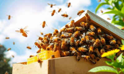
“Revolutionizing Honey Bee Survival: A New Pollen-Replacing Food Source Brings Hope for the Future”