Connect with us

Detectors

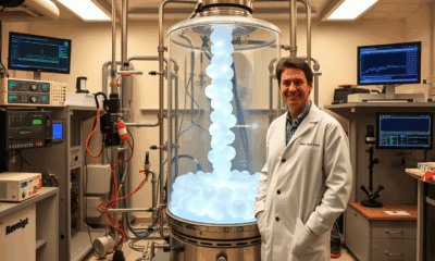
Revolutionizing Industrial Imaging with Laser-Driven CT Scans

A research team has achieved a new milestone in 3D X-ray imaging technology. The scientists have captured high-resolution CT scans of the interior of a large, dense object — a gas turbine blade — using a compact, laser-driven X-ray source. The work is part of a larger vision to leverage high-intensity lasers for a wide range of uses, from studying inertial fusion energy to generating bright beams of GeV electrons and MeV x-rays.
Avatar photo

Published

on

The world of industrial imaging has just taken a giant leap forward. Researchers at Colorado State University have successfully captured high-resolution CT scans of dense objects, including a large gas turbine blade, using a compact, laser-driven X-ray source. This breakthrough achievement, published in Optica, marks the beginning of a new era in radiographic imaging technology.

The research team, led by assistant professor Reed Hollinger, has been working tirelessly for years to perfect this innovative approach. By leveraging the CSU-built ALEPH laser, they have generated extremely bright X-ray sources capable of producing high-resolution images without compromising energy levels. This significant advancement opens doors to various industries, including aerospace and additive manufacturing.

The implications are vast. With this technology, manufacturers can obtain a detailed view inside dense structures, such as rocket components and turbojet engines, using a fast and non-destructive method. Additive manufacturing could benefit greatly from quality control enhancements, preserving the integrity of 3D-printed parts.

Current industrial CT scanners are massive and costly, producing images with millimeter-scale resolution. In contrast, the laser-driven approach generates a much smaller X-ray source, enabling significantly higher resolution without decreasing energy levels. This is made possible by accelerating electrons to millions of volts using a petawatt-class laser, which then collide with heavy atoms to produce high-energy X-rays.

The increased X-ray energy is essential for penetrating dense objects like the turbine blade shown in the study. Each X-ray pulse only lasts for a few trillionths of a second, allowing time-resolved radiography of objects moving at incredible speeds.

Imagine being able to capture high-resolution 3D images of the inside of a jet engine while it’s operating – currently an impossible feat with existing X-ray sources. This is the kind of revolutionary imaging that researchers aim to achieve using the expanded capabilities of CSU’s Advanced Technology Lasers for Applications and Science (ATLAS) Facility, set to come online in late 2026.

The possibilities are endless, and the future of industrial imaging has never looked brighter. With this laser-driven CT scan technology, manufacturers can unlock new levels of quality control, efficiency, and innovation – all while preserving the integrity of their products.

Detectors

Green fabrication of hybrid materials as highly sensitive X-ray detectors: A breakthrough for medical diagnostics and material characterization.

New bismuth-based organic-inorganic hybrid materials show exceptional sensitivity and long-term stability as X-ray detectors, significantly more sensitive than commercial X-ray detectors. In addition, these materials can be produced without solvents by ball milling, a mechanochemical synthesis process that is environmentally friendly and scalable. More sensitive detectors would allow for a reduction in the radiation exposure during X-ray examinations.

Avatar photo

Published

on

New bismuth-based organic-inorganic hybrid materials have been discovered, showing exceptional sensitivity and long-term stability as X-ray detectors. This breakthrough is set to transform the field of medical diagnostics and material characterization, allowing for a significant reduction in radiation exposure during X-ray examinations.

X-ray imaging is an indispensable tool in various fields, including medicine and material science. To generate accurate images, high-quality detectors are essential. Current commercial detectors consist of inorganic compounds with medium to high atomic numbers. However, these materials have limitations, particularly in terms of sensitivity and stability.

The development of new bismuth-based organic-inorganic hybrid materials has addressed these limitations. Inspired by the success of halide perovskite compounds in opto-electronic devices, researchers at HZB have created two novel materials: [(CH3CH2)3S]6Bi8I30 and [(CH3CH2)3S]AgBiI5. These materials demonstrate exceptional sensitivity and stability, making them ideal for X-ray detection.

A particularly environmentally friendly manufacturing process was used to produce these materials: ball milling. This method produces polycrystalline powders that are then pressed into dense pellets. The procedures involved are also established in industry, making the production of these hybrid materials scalable and sustainable.

The novel materials were evaluated for their use in X-ray detectors, with impressive results. They show sensitivities up to two orders of magnitude higher than commercial materials like amorphous selenium or CdZnTe – and can detect X-ray doses nearly 50 times lower.

The team also studied the samples at the KMC-3 XPP beamline at BESSY II, where the detectors maintained a stable response during pulsed X-ray irradiation under high-intensity photon flux. No measurable degradation in performance was observed post-exposure, highlighting the robustness of the detector materials.

This breakthrough has significant implications for medical diagnostics and material characterization. The development of highly sensitive X-ray detectors using bismuth-based organic-inorganic hybrid materials has the potential to reduce radiation exposure during X-ray imaging. This is particularly important in medical applications, where minimizing radiation exposure is crucial.

The next step is technology transfer, with opportunities to collaborate with companies in Adlershof to optimize the development of such X-ray detectors. This collaboration will enable the translation of this scientific breakthrough into practical applications, revolutionizing the field of medical diagnostics and material characterization.

Continue Reading

Astrophysics

“Revolutionizing Mid-Infrared Detection: KAIST Researchers Develop Groundbreaking Photodetector for Exoplanet Hunting and Beyond”

Researchers have developed an innovative photodetector capable of detecting a broad range of mid-infrared spectra.

Avatar photo

Published

on

By

In a groundbreaking innovation, researchers from Korea Advanced Institute of Science and Technology (KAIST) have developed a mid-infrared photodetector that can detect a broad range of spectral signals at room temperature. This breakthrough has significant implications for exoplanet hunting, environmental monitoring, medical diagnostics, and other fields.

The new photodetector, led by Professor SangHyeon Kim from the School of Electrical Engineering, utilizes conventional silicon-based CMOS processes, enabling low-cost mass production while maintaining stable operation at room temperature. This is a major departure from existing mid-infrared photodetectors, which typically require cooling systems and are incompatible with silicon-based CMOS processes.

The research team successfully demonstrated the real-time detection of carbon dioxide (CO2) gas using ultra-compact and ultra-thin optical sensors equipped with this photodetector. This has proven its potential for environmental monitoring and hazardous gas analysis. Moreover, the waveguide-integrated design of this new technology allows it to detect the entire mid-infrared spectral range, making it suitable for real-time sensing of various molecular species.

The development of this groundbreaking photodetector overcomes the limitations of existing mid-infrared sensor technologies, including the need for cooling systems, difficulties in mass production, and high costs. This breakthrough technology is expected to be applicable across diverse fields, including environmental monitoring, medical diagnostics, industrial process management, national defense and security, and smart devices.

According to Professor Kim, “This research represents a novel approach that overcomes the limitations of existing mid-infrared photodetector technologies and has great potential for practical applications in various fields.” He further emphasized, “Since this sensor technology is compatible with CMOS processes, it enables low-cost mass production, making it highly suitable for next-generation environmental monitoring systems and smart manufacturing sites.”

The study was published on March 19, 2025, in the journal Light: Science & Applications. This innovative technology has the potential to revolutionize various fields and pave the way for next-generation mid-infrared sensor advancements.

Continue Reading

Biotechnology

Breaking Ground: Ultrasound Revolutionizes Hydrogel Manufacturing

Researchers have developed a new way to create hydrogels using ultrasound, eliminating the need for toxic chemical initiators. This breakthrough offers a faster, cleaner and more sustainable approach to hydrogel fabrication, and produces hydrogels that are stronger, more flexible and highly resistant to freezing and dehydration. The new method also promises to facilitate advances in tissue engineering, bioadhesives and 3D bioprinting.

Avatar photo

Published

on

The world of hydrogel manufacturing has just gotten a whole lot greener. Researchers at McGill University, in collaboration with Polytechnique Montréal, have pioneered a groundbreaking method to create hydrogels using ultrasound, eliminating the need for toxic chemical initiators. This innovation promises a faster, cleaner, and more sustainable approach to hydrogel fabrication, producing materials that are stronger, more flexible, and highly resistant to freezing and dehydration.

Hydrogels, composed of polymers that can absorb and retain large amounts of water, have numerous applications in wound dressings, drug delivery, tissue engineering, soft robotics, and more. Traditional hydrogel manufacturing relies on chemical initiators, some of which can be hazardous, particularly in medical applications. These chemicals trigger chemical chain reactions, but the McGill research team has developed an alternative method using ultrasound.

When applied to a liquid precursor, sound waves create microscopic bubbles that collapse with immense energy, triggering gel formation within minutes. This ultrasound-driven technique is dubbed “sonogel.” According to Mechanical Engineering Professor Jianyu Li, who led the research team, the problem they aimed to solve was the reliance on toxic chemical initiators.

“Our method eliminates these substances, making the process safer for the body and better for the environment,” said Li. With sonogel, gel formation occurs in just five minutes, compared to hours or even overnight under UV light. This speed and efficiency have significant implications for biomedical applications.

One of the most exciting possibilities for this technology is in non-invasive medical treatments. Because ultrasound waves can penetrate deep into tissues, this method could enable in-body hydrogel formation without surgery. Imagine injecting a liquid precursor and using ultrasound to solidify it precisely where needed – this could be a game-changer for treating tissue damage and regenerative medicine.

Further refinement of this technique also opens the door to ultrasound-based 3D bioprinting. Instead of relying on light or heat, researchers could use sound waves to precisely “print” hydrogel structures. By leveraging high-intensity focused ultrasound, researchers can shape and build hydrogels with remarkable precision.

According to Jean Provost, one of co-authors of the study and assistant professor of engineering physics at Polytechnique Montréal, this breakthrough has significant potential for safer, greener material production. The sonogel method has the potential to revolutionize biomedical applications and unlock new possibilities for non-invasive medical treatments, making it a truly groundbreaking innovation in the field of hydrogel manufacturing.

Continue Reading

Trending