Connect with us
We’re experimenting with AI-generated content to help deliver information faster and more efficiently.
While we try to keep things accurate, this content is part of an ongoing experiment and may not always be reliable.
Please double-check important details — we’re not responsible for how the information is used.

Brain Injury

“Revolutionizing Stroke Recovery: A Breakthrough Drug that Repairs Brain Damage”

A new study has discovered what researchers say is the first drug to fully reproduce the effects of physical stroke rehabilitation in model mice, following from human studies.
Avatar photo

Published

on

The article you provided highlights a groundbreaking study by UCLA Health that has identified the first drug to fully replicate the effects of physical stroke rehabilitation in model mice. This breakthrough discovery, published in Nature Communications, has significant implications for the treatment of stroke patients.

Stroke is the leading cause of adult disability, with most patients not fully recovering from its effects. Currently, there are no drugs available to aid in stroke recovery, requiring patients to undergo physical rehabilitation, which has shown limited effectiveness. Dr. S. Thomas Carmichael, the study’s lead author and professor and chair of UCLA Neurology, emphasizes the need for a medicine that can produce the same benefits as rehabilitation.

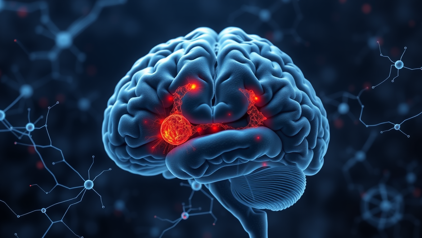
The researchers sought to determine how physical rehabilitation improved brain function after a stroke and whether they could generate a drug that could replicate these effects. They identified a loss of brain connections in mice models of stroke, which led to disconnection between neurons, resulting in impaired movement and gait. The team found that the loss of connections occurred in parvalbumin neurons, which help generate gamma oscillations – a brain rhythm essential for coordinated networks.

The researchers then developed two candidate drugs that specifically target parvalbumin neurons to excite them and restore gamma oscillations. One of these drugs, DDL-920, showed significant recovery in movement control in mouse models. While further studies are needed to understand the safety and efficacy of this drug before it can be considered for human trials, this breakthrough offers new hope for stroke patients and their families.

The discovery of a drug that can repair brain damage caused by stroke has the potential to revolutionize the treatment of this condition, allowing patients to recover more fully and regain control over their lives.

Brain Injury

Unveiling the Hidden Brain Network for Naming

Researchers identified two brain networks involved in word retrieval — the cognitive process of accessing words we need to speak. A semantic network processes meaning in middle/inferior frontal gyri, while an articulatory network in inferior frontal/precentral gyri plans speech production.

Avatar photo

Published

on

By

The human brain is capable of performing incredible feats, from recalling memories to navigating complex mathematical equations. Yet, there lies one basic yet essential ability that often goes unnoticed – the power to name words we want to say. This seemingly simple act, called word retrieval, can be severely compromised in individuals with brain damage or neurological disorders. Despite decades of research, scientists have long sought to understand how the brain retrieves words during speech.

A groundbreaking study by researchers at New York University has shed light on this mystery, revealing a left-lateralized network in the dorsolateral prefrontal cortex that plays a crucial role in naming. Published in Cell Reports, the findings provide new insights into the neural architecture of language, with potential applications for both neuroscience and clinical interventions.

The study involved recording electrocorticographic (ECoG) data from 48 neurosurgical patients to examine the spatial and temporal organization of language processing in the brain. By using unsupervised clustering techniques, the researchers identified two distinct but overlapping networks responsible for word retrieval – a semantic processing network located in the middle and inferior frontal gyri, and an articulatory planning network situated in the inferior frontal and precentral gyri.

A striking ventral-dorsal gradient was observed in the prefrontal cortex, with articulatory planning localized ventrally and semantic processing uniquely represented in a dorsal region of the inferior frontal gyrus and middle frontal gyrus. This previously underappreciated hub for language processing has been found to play a crucial role in mapping sounds to meaning in an auditory context.

The findings have far-reaching implications, not only for theoretical neuroscience but also for clinical applications. Language deficits, such as anomia – the inability to retrieve words – are common in stroke, brain injury, and neurodegenerative disorders. Understanding the precise neural networks involved in word retrieval could lead to better diagnostics and targeted rehabilitation therapies for patients suffering from these conditions.

Additionally, the study provides a roadmap for future research in brain-computer interfaces (BCIs) and neuroprosthetics. By decoding the neural signals associated with naming, scientists could potentially develop assistive devices for individuals with speech impairments, allowing them to communicate more effectively through direct brain-computer communication.

In conclusion, our ability to name the world around us is not just a simple act of recall but the result of a sophisticated and finely tuned neural system – one that is now being revealed in greater detail than ever before. The discovery of this hidden brain network has opened up new avenues for research and potential applications, ultimately improving our understanding of human language and cognition.

Continue Reading

Birth Defects

Unconsciousness by Design: How Anesthetics Shift Brainwave Phase to Induce Slumber

A new study finds that an easily measurable brain wave shift of phase may be a universal marker of unconsciousness under general anesthesia.

Avatar photo

Published

on

The rewritten article aims to make the complex scientific concepts more accessible to a general audience while maintaining the core ideas and findings of the original study.

Unconsciousness by Design: How Anesthetics Shift Brainwave Phase to Induce Slumber

Scientists have long been fascinated by the mysterious world of unconsciousness, trying to understand what happens in our brains when we fall asleep or are anesthetized. A new study has shed light on this phenomenon, revealing a common thread among different anesthetics: they all induce unconsciousness by shifting brainwave phase.

Ketamine and dexmedetomidine, two distinct anesthetics with different molecular mechanisms, were used in the study to demonstrate how these drugs achieve the same result – inducing unconsciousness. By analyzing brain wave activity, researchers found that both anesthetics push around brain waves, causing them to fall out of phase.

In a conscious state, local groups of neurons in the brain’s cortex can share information to produce cognitive functions such as attention, perception, and reasoning. However, when brain waves become misaligned, these local communications break down, leading to unconsciousness.

The study, led by graduate student Alexandra Bardon, discovered that the way anesthetics shift brainwave phase is a potential signature of unconsciousness that can be measured. This finding has significant implications for anesthesiology care, as it could provide a common new measure for anesthesiologists to ensure patients remain unconscious during surgery.

“If you look at the way phase is shifted in our recordings, you can barely tell which drug it was,” said Earl K. Miller, senior author of the study and Picower Professor. “That’s valuable for medical practice.”

The researchers also found that distance played a crucial role in determining the change in phase alignment. Even across short distances, low-frequency waves moved out of alignment, with a 180-degree shift observed between arrays in the upper and lower regions within a hemisphere.

This study raises many opportunities for follow-up research, including exploring how other anesthetics affect brainwave phase and investigating the role of traveling waves in the phenomenon. Furthermore, understanding the difference between anesthesia-induced unconsciousness and sleep could lead to new insights into the mechanisms that generate consciousness.

In conclusion, this study provides a fascinating glimpse into the world of unconsciousness, revealing a common thread among different anesthetics. By continuing to explore the intricacies of brainwave phase alignment, scientists may uncover more secrets about the mysteries of the human brain.

Continue Reading

Accident and Trauma

Lowered Tackle Height Reduces Head Contacts in Women’s Rugby, Study Finds

Lowering the legal tackle height in women’s rugby is proving effective in reducing head contacts between players, a new study suggests. Changes to the tackle height law in women’s community rugby in Scotland is linked to reductions in head-to-head and head-to shoulder contacts, the study found. The researchers used video analysis to study the impact of the lowered tackle height law which World Rugby, the sport’s governing body, introduced for community rugby in an attempt to improve safety for players.

Avatar photo

Published

on

The lowered legal tackle height in women’s community rugby has shown to be effective in reducing head contacts between players, according to a world-first study published in BMJ Open Sport and Exercise Medicine. The research, conducted by researchers at the University of Edinburgh in collaboration with Scottish Rugby and World Rugby, analyzed video footage from 34 Scottish community women’s rugby matches played before and after the introduction of the lowered tackle height law.

The study found that the reduced tackle height led to a significant decrease in head-to-head and head-to-shoulder contacts between players. Specifically, the research revealed:

* A 21% reduction in upright tackles
* A 34% increase in tacklers entering the tackle bent at the waist, which is considered the recommended technique to reduce contact with high-risk areas of the head and shoulders
* A 64% reduction in tacklers making initial contact with the ball carrier’s head and neck
* A 17% reduction in the rate of head-to-head contacts for the tackler
* A 35% reduction in head-to-shoulder contacts for the tackler

The study also found a 19% reduction in contacts above the sternum, known as the “red zone,” between the tackler and the ball carrier. This is considered an area of high risk for concussion.

While the study did not find a significant change in the rate of concussions and injuries when comparing the pre-trial and trial seasons, researchers note that the number of reported injuries overall was very low and may have impacted these findings.

The lead author of the study, Hannah Walton from the University of Edinburgh’s Moray House School of Education and Sport, emphasized the importance of continued collection of robust tackle and injury data to further understand the effect of the law change on player behavior and safety.

This research provides valuable insights into the impact of lowering the tackle height in women’s community rugby and can inform future injury prevention initiatives. The study is part of an international project led by World Rugby to assess the effects of lowering the tackle height in 11 countries, including Australia, England, France, Ireland, Italy, Japan, New Zealand, Scotland, South Africa, and Wales.

Continue Reading

Trending