Connect with us
We’re experimenting with AI-generated content to help deliver information faster and more efficiently.
While we try to keep things accurate, this content is part of an ongoing experiment and may not always be reliable.
Please double-check important details — we’re not responsible for how the information is used.

Biochemistry Research

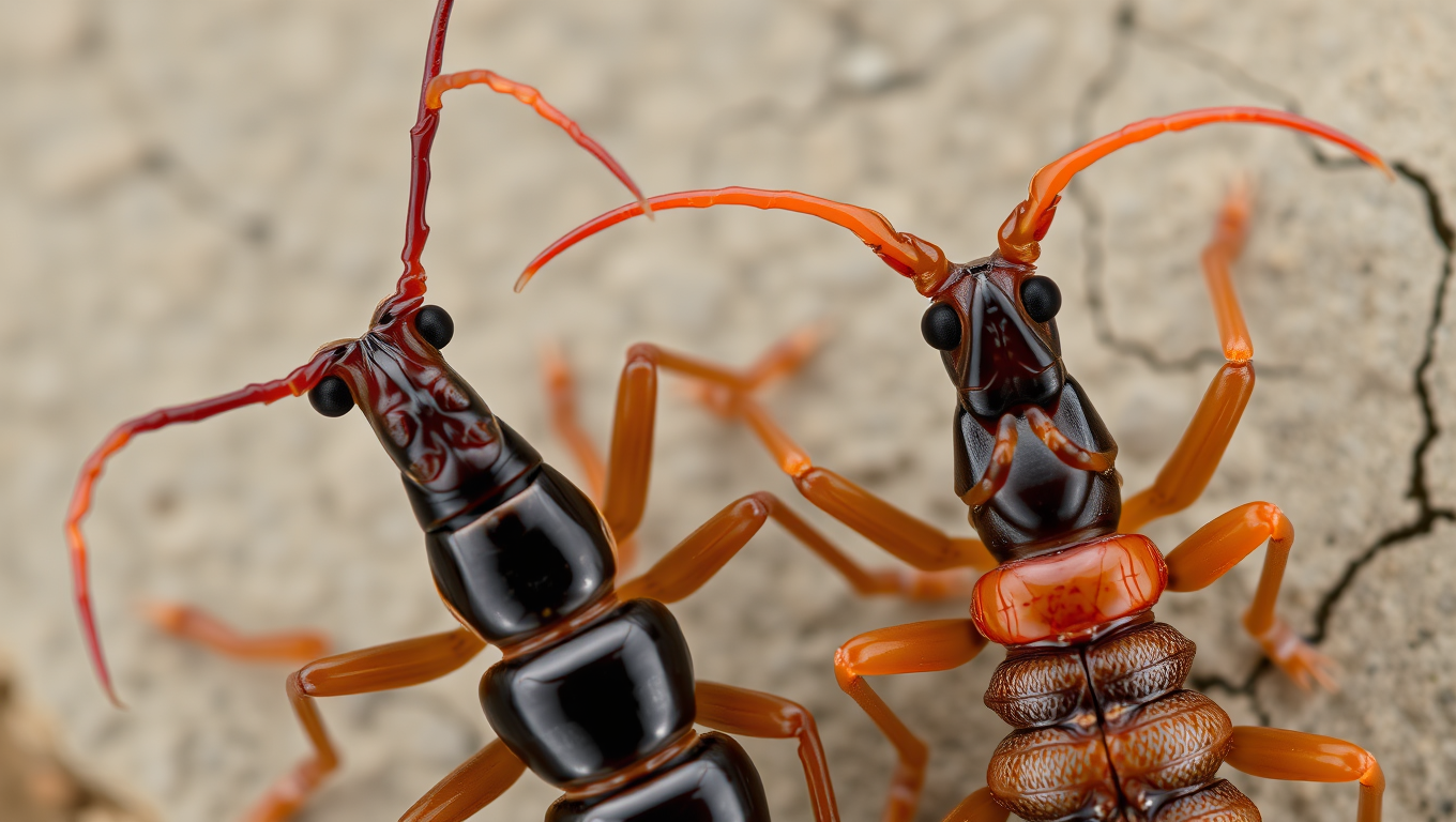
The Double Edge of Love and War: How Female Earwigs Evolved Deadly Claws for Mate Competition

Female earwigs may be evolving exaggerated weaponry just like males. A study from Toho University found that female forceps, once assumed to be passive tools, show the same kind of outsized growth linked to sexual selection as the male’s iconic pincers. This means that female earwigs might be fighting for mates too specifically for access to non-aggressive males challenging long-standing assumptions in evolutionary biology.
Avatar photo

Published

on

The article you provided has been rewritten to improve clarity, structure, and style while maintaining its core ideas. Here is the rewritten content:

In a groundbreaking study published in the Biological Journal of the Linnean Society on June 12, 2025, researchers from Toho University have shed new light on the evolution of deadly claws in female earwigs. For decades, it was believed that these pincer-like appendages were exclusive to males and evolved solely as weapons in battles with rivals. However, the findings of Tomoki Matsuzawa (then an undergraduate) and Associate Professor Junji Konuma have challenged this notion, revealing a surprising parallel between male and female earwigs.

The researchers conducted a quantitative study on the maritime earwig Anisolabis maritima, analyzing the morphometric data of both sexes. They found that not only do females possess forceps, but they also exhibit positive allometry – a phenomenon where certain body parts grow disproportionately large relative to body size. This is strikingly similar to the pattern observed in males, suggesting that female earwigs may have evolved these traits through sexual selection.

In their study, the team measured various dimensions of the head, thorax, abdomen, and bilateral forceps, as well as shape differences between sexes. They discovered that males possess thick, short, and curved forceps, while females have thin, long, and straight ones – a clear example of sexual dimorphism. When they plotted body size against forceps width and length on a log-log scale, the results revealed positive allometry in both males (in forceps width) and females (in forceps length).

Associate Professor Konuma explained that this finding suggests female earwigs may have evolved their forceps as effective weapons in competing for mates. A previous behavioral study had shown that female earwigs engage in competition with each other for small, non-aggressive males. This new research highlights the importance of considering female traits when studying the evolution of insect morphologies.

These groundbreaking findings demonstrate how the complex and fascinating world of insects can continue to surprise us, revealing the intricacies of natural selection and mate competition.

Biochemistry Research

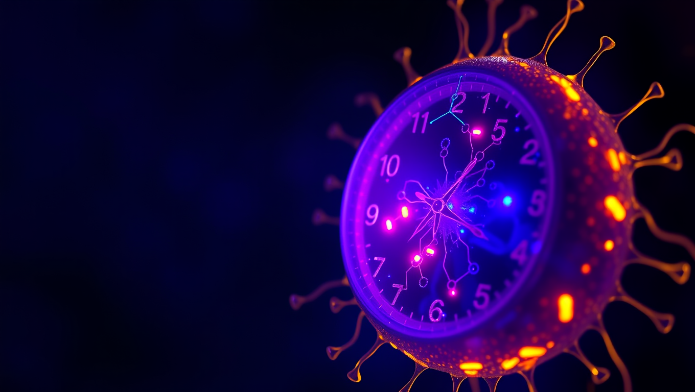
“Unlocking Timekeeping Secrets: Scientists Reveal How Artificial Cells Can Accurately Keep Rhythm”

Scientists at UC Merced have engineered artificial cells that can keep perfect time—mimicking the 24-hour biological clocks found in living organisms. By reconstructing circadian machinery inside tiny vesicles, the researchers showed that even simplified synthetic systems can glow with a daily rhythm—if they have enough of the right proteins.

Avatar photo

Published

on

A team of researchers from UC Merced has made a groundbreaking discovery by creating tiny artificial cells that can accurately keep time, mimicking the daily rhythms found in living organisms. This achievement sheds light on how biological clocks stay on schedule despite the inherent molecular noise inside cells.

The study, published in Nature Communications, was led by bioengineering Professor Anand Bala Subramaniam and chemistry and biochemistry Professor Andy LiWang. The team’s findings show that artificial cells can glow in a regular 24-hour rhythm for at least four days when loaded with core clock proteins, one of which is tagged with a fluorescent marker.

However, when the number of clock proteins is reduced or the vesicles are made smaller, the rhythmic glow stops. This loss of rhythm follows a reproducible pattern, indicating that clocks become more robust with higher concentrations of clock proteins, allowing thousands of vesicles to keep time reliably – even when protein amounts vary slightly between vesicles.

To explain these findings, the team built a computational model that revealed another component of the natural circadian system – responsible for turning genes on and off – does not play a major role in maintaining individual clocks but is essential for synchronizing clock timing across a population. The researchers also noted that some clock proteins tend to stick to the walls of the vesicles, meaning a high total protein count is necessary to maintain proper function.

“This study shows that we can dissect and understand the core principles of biological timekeeping using simplified, synthetic systems,” Subramaniam said.

The work led by Subramaniam and LiWang advances the methodology for studying biological clocks, according to Mingxu Fang, a microbiology professor at Ohio State University and an expert in circadian clocks. “This new study introduces a method to observe reconstituted clock reactions within size-adjustable vesicles that mimic cellular dimensions,” Fang said. “This powerful tool enables direct testing of how and why organisms with different cell sizes may adopt distinct timing strategies, thereby deepening our understanding of biological timekeeping mechanisms across life forms.”

The study was supported by Subramaniam’s National Science Foundation CAREER award from the Division of Materials Research and by grants from the National Institutes of Health and Army Research Office awarded to LiWang.

Continue Reading

Behavioral Science

The Sugar that Sparked Life: Unraveling the Mystery of Ribose’s Preeminence in RNA Development

What made ribose the sugar of choice for life’s code? Scientists at Scripps Research may have cracked a major part of this mystery. Their experiments show that ribose binds more readily and selectively to phosphate compared to other similar sugars, forming a structure ideal for RNA formation. This discovery hints at how nature might have selected specific molecules long before enzymes or life existed, and could reshape our understanding of life’s chemical origins.

Avatar photo

Published

on

The study published in Angewandte Chemie sheds light on how ribose may have become the preferred sugar for RNA development, highlighting its unique ability to bind with phosphate more quickly and effectively than other sugar molecules. This characteristic could have played a crucial role in selecting ribose as the building block of life.

Ramanarayanan Krishnamurthy, professor of chemistry at Scripps Research, emphasizes that this finding supports the idea that prebiotic chemistry could have produced the fundamental components of RNA, which eventually led to entities exhibiting lifelike properties. The research focuses on phosphorylation, a step within nucleotide-building where ribose connects to the phosphate group, and explores whether other sugars can undergo similar reactions.

The team’s experiments showed that while diamidophosphate (DAP) could phosphorylate all four sugar molecules tested, it phosphorylated ribose at a significantly faster rate. The reaction with ribose produced exclusively ring-shaped structures with five corners, whereas the other sugars formed a combination of 5- and 6-member rings.

“This really showed us that there is a difference between ribose and the three other sugars,” says Krishnamurthy. “Ribose not only reacts faster than the other sugars, it’s also more selective for the five-member ring form, which happens to be the form that we see in RNA and DNA today.”

When DAP was added to a solution containing equal amounts of the four different sugars, it preferentially phosphorylated ribose. The researchers demonstrated that this selective process produces a molecule with a form conducive for making RNA, providing further evidence for ribose’s preeminence.

While the study does not claim that these reactions directly led to life, it suggests that they might have played a crucial role in the primordial process that gave rise to the fundamental components of life. The researchers caution against over-interpretation and emphasize the need for further investigation into the emergence of life on Earth.

In future research, the team plans to test whether this chemical reaction can occur inside primitive cellular structures called protocells. If successful, it might provide a compelling explanation for how ribose became the preferred sugar for RNA development and ultimately gave rise to life as we know it today.

Continue Reading

Biochemistry Research

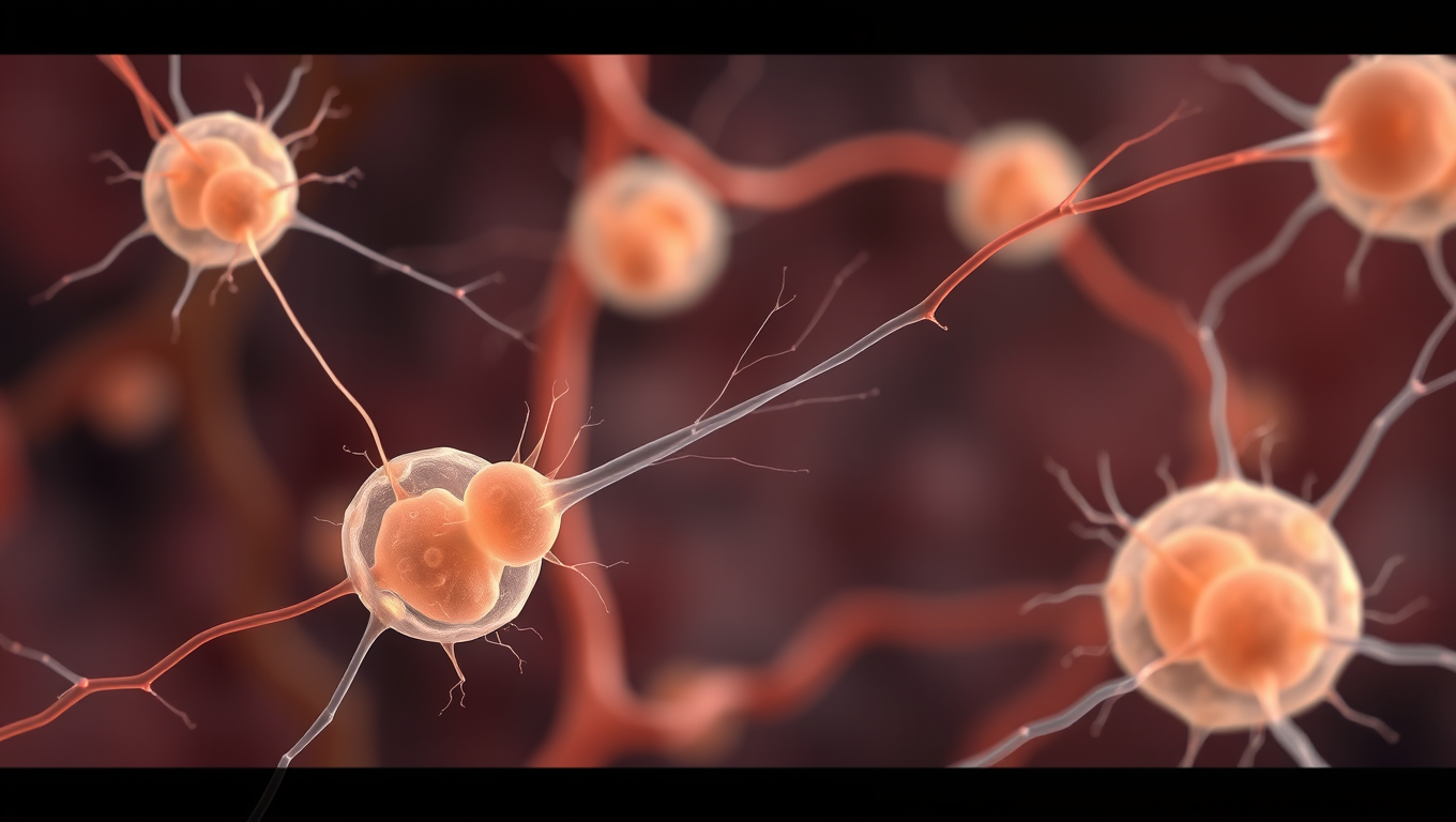
The Whispering Womb: Uncovering the Secret Language of Embryonic Cells

Scientists found that embryonic skin cells “whisper” through faint mechanical tugs, using the same force-sensing proteins that make our ears ultrasensitive. By syncing these micro-movements, the cells choreograph the embryo’s shape, a dance captured with AI-powered imaging and computer models. Blocking the cells’ ability to feel the whispers stalls development, hinting that life’s first instructions are mechanical. The discovery suggests hearing hijacked an ancient force-sensing toolkit originally meant for building bodies.

Avatar photo

Published

on

The human body begins as a single cell that multiplies and differentiates into thousands of specialized cells. Researchers at the Göttingen Campus Institute for Dynamics of Biological Networks (CIDBN) and the Max Planck Institute have made a groundbreaking discovery: embryonic cells “listen” to each other through molecular mechanisms previously known only from hearing.

Using an interdisciplinary approach combining developmental genetics, brain research, hearing research, and theoretical physics, the researchers found that in thin layers of skin, cells register the movements of their neighboring cells and synchronize their own tiny movements with those of the others. This coordination allows groups of neighboring cells to pull together with greater force, making them highly sensitive and able to respond quickly and flexibly.

The researchers created computer models of tissue development, which showed that this “whispering” among neighboring cells leads to an intricate choreography of the entire tissue, protecting it from external forces. These findings were confirmed by video recordings of embryonic development and further experiments.

Dr. Matthias Häring, group leader at the CIDBN, explained that using AI methods and computer-assisted analysis allowed them to examine about a hundred times more cell pairs than was previously possible in this field, giving their results high accuracy.

The mechanisms revealed in embryonic development are also known to play a role in hearing, where hair cells convert sound waves into nerve signals. The ear is sensitive because of special proteins that convert mechanical forces into electrical currents. This discovery suggests that such sensors of force may have evolved from our single-celled ancestors, which emerged long before the origin of animal life.

Professor Fred Wolf, Director of the CIDBN, noted that future work should determine whether the original function of these cellular “nanomachines” was to perceive forces inside the body rather than perceiving the outside world. This phenomenon could provide insights into how force perception at a cellular level has evolved.

Continue Reading

Trending