While we try to keep things accurate, this content is part of an ongoing experiment and may not always be reliable.
Please double-check important details — we’re not responsible for how the information is used.
Cancer
The Next-Generation Radiation Detector: A Game-Changer for Safety and Security
Scientists have developed a new type of handheld multi-purpose radiation detector that comprehensively detects all types of ionizing radiation. The device can be used by industrial and medical radiation users, regulatory authorities, the nuclear energy industry, first responders and military users. The technology has been patented and is currently being explored for commercialization.


Cancer
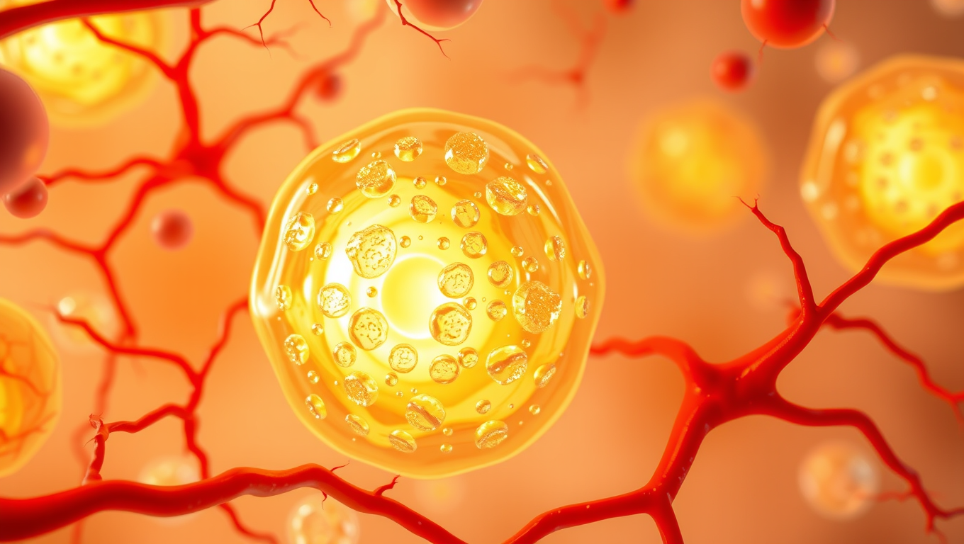
A Silent Killer Unmasked: The Hidden Gene in Leukemia Virus that Could Revolutionize HIV Treatment
Scientists in Japan have discovered a genetic “silencer” within the HTLV-1 virus that helps it stay hidden in the body, evading the immune system for decades. This silencer element essentially turns the virus off, preventing it from triggering symptoms in most carriers. Incredibly, when this silencer was added to HIV, it made that virus less active too — hinting at a revolutionary new strategy for managing not just HTLV-1 but other deadly retroviruses as well. The discovery opens the door to turning the virus’s own stealth tactics against it in future treatments.

Cancer
Turning Yogurt into a Healing Gel: Columbia Scientists Pioneer New Regenerative Medicine Approach
Scientists at Columbia Engineering have developed an injectable hydrogel made from yogurt-derived extracellular vesicles (EVs) that could revolutionize regenerative medicine. These EVs serve both as healing agents and as structural components, eliminating the need for added chemicals. The innovation leverages everyday dairy products like yogurt to create a biocompatible material that mimics natural tissue and enhances healing.
Cancer
Safer Non-Stick Coatings: Scientists Develop Alternative to Teflon
Scientists at the University of Toronto have developed a new non-stick material that rivals the performance of traditional PFAS-based coatings while using only minimal amounts of these controversial “forever chemicals.” Through an inventive process called “nanoscale fletching,” they modified silicone-based polymers to repel both water and oil effectively. This breakthrough could pave the way for safer cookware, fabrics, and other products without the environmental and health risks linked to long-chain PFAS.


A New Horizon for Vision: How Gold Nanoparticles May Restore People’s Sight

Revolutionizing Quantum Communication: Direct Connections Between Multiple Processors

Retiring Abroad Can Be Lonely Business

“Unveiling Hidden Patterns: A New Twist on Interference Phenomena”
Harnessing Water Waves: A Breakthrough in Controlling Floating Objects

Household Electricity Three Times More Expensive Than Upcoming ‘Eco-Friendly’ Aviation E-Fuels, Study Reveals
Reducing Falls Among Elderly Women with Polypharmacy through Exercise Intervention
-
 Detectors1 year ago
A New Horizon for Vision: How Gold Nanoparticles May Restore People’s Sight
-
Cancer1 year ago
Revolutionizing Quantum Communication: Direct Connections Between Multiple Processors
-
Earth & Climate1 year ago
Retiring Abroad Can Be Lonely Business
-
Chemistry1 year ago
“Unveiling Hidden Patterns: A New Twist on Interference Phenomena”
-
Albert Einstein1 year ago
Harnessing Water Waves: A Breakthrough in Controlling Floating Objects
-
Earth & Climate1 year ago
Household Electricity Three Times More Expensive Than Upcoming ‘Eco-Friendly’ Aviation E-Fuels, Study Reveals
-
Diseases and Conditions1 year ago
Reducing Falls Among Elderly Women with Polypharmacy through Exercise Intervention
-
Acid Rain1 year ago
“Revolutionizing Honey Bee Survival: A New Pollen-Replacing Food Source Brings Hope for the Future”