While we try to keep things accurate, this content is part of an ongoing experiment and may not always be reliable.
Please double-check important details — we’re not responsible for how the information is used.
Alzheimer's Research
The Rise of Dry Eye Disease in Young Adults: A Growing Concern
Researchers have called for more advice to be given to young people about preventing dry eye disease, after a study found that 90% of participants had at least one sign of the condition in their eyes.


Alzheimer's
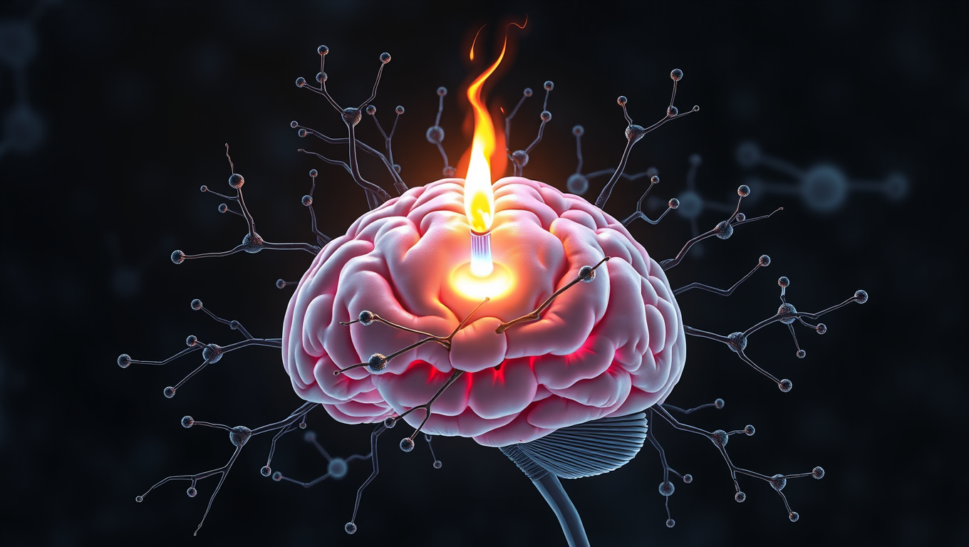
Scientists Unlock Secret to Reversing Memory Loss by Boosting Brain’s Energy Engines
Scientists have discovered a direct cause-and-effect link between faulty mitochondria and the memory loss seen in neurodegenerative diseases. By creating a novel tool to boost mitochondrial activity in mouse models, researchers restored memory performance, suggesting mitochondria could be a powerful new target for treatments. The findings not only shed light on the early drivers of brain cell degeneration but also open possibilities for slowing or even preventing diseases like Alzheimer’s.
Alzheimer's
A Breakthrough in Brain Research: Scientists Grow a Mini Human Brain that Lights Up and Connects Like the Real Thing
Scientists at Johns Hopkins have grown a first-of-its-kind organoid mimicking an entire human brain, complete with rudimentary blood vessels and neural activity. This new “multi-region brain organoid” connects different brain parts, producing electrical signals and simulating early brain development. By watching these mini-brains evolve, researchers hope to uncover how conditions like autism or schizophrenia arise, and even test treatments in ways never before possible with animal models.
Alzheimer's
“Unlocking Brain Health: Scientists Discover Key Receptor for Microglia to Fight Alzheimer’s”
Scientists at UCSF have uncovered how certain immune cells in the brain, called microglia, can effectively digest toxic amyloid beta plaques that cause Alzheimer’s. They identified a key receptor, ADGRG1, that enables this protective action. When microglia lack this receptor, plaque builds up quickly, causing memory loss and brain damage. But when the receptor is present, it seems to help keep Alzheimer’s symptoms mild. Since ADGRG1 belongs to a drug-friendly family of receptors, this opens the door to future therapies that could enhance brain immunity and protect against Alzheimer’s in more people.


A New Horizon for Vision: How Gold Nanoparticles May Restore People’s Sight

Revolutionizing Quantum Communication: Direct Connections Between Multiple Processors

Retiring Abroad Can Be Lonely Business
Harnessing Water Waves: A Breakthrough in Controlling Floating Objects

Household Electricity Three Times More Expensive Than Upcoming ‘Eco-Friendly’ Aviation E-Fuels, Study Reveals

“Unveiling Hidden Patterns: A New Twist on Interference Phenomena”
Reducing Falls Among Elderly Women with Polypharmacy through Exercise Intervention
-
 Detectors1 year ago
A New Horizon for Vision: How Gold Nanoparticles May Restore People’s Sight
-
Cancer1 year ago
Revolutionizing Quantum Communication: Direct Connections Between Multiple Processors
-
Earth & Climate1 year ago
Retiring Abroad Can Be Lonely Business
-
Albert Einstein1 year ago
Harnessing Water Waves: A Breakthrough in Controlling Floating Objects
-
Earth & Climate1 year ago
Household Electricity Three Times More Expensive Than Upcoming ‘Eco-Friendly’ Aviation E-Fuels, Study Reveals
-
Chemistry1 year ago
“Unveiling Hidden Patterns: A New Twist on Interference Phenomena”
-
Diseases and Conditions1 year ago
Reducing Falls Among Elderly Women with Polypharmacy through Exercise Intervention
-
Earth & Climate1 year ago
Predicting Damage from Local Earthquakes in Mexico City