
While we try to keep things accurate, this content is part of an ongoing experiment and may not always be reliable.
Please double-check important details — we’re not responsible for how the information is used.
Breast Cancer
The Surprising Link Between Diet, Gut Microbes, and Cancer Therapy Efficacy
A study has uncovered a surprising link between diet, intestinal microbes and the efficacy of cancer therapy.


Breast Cancer
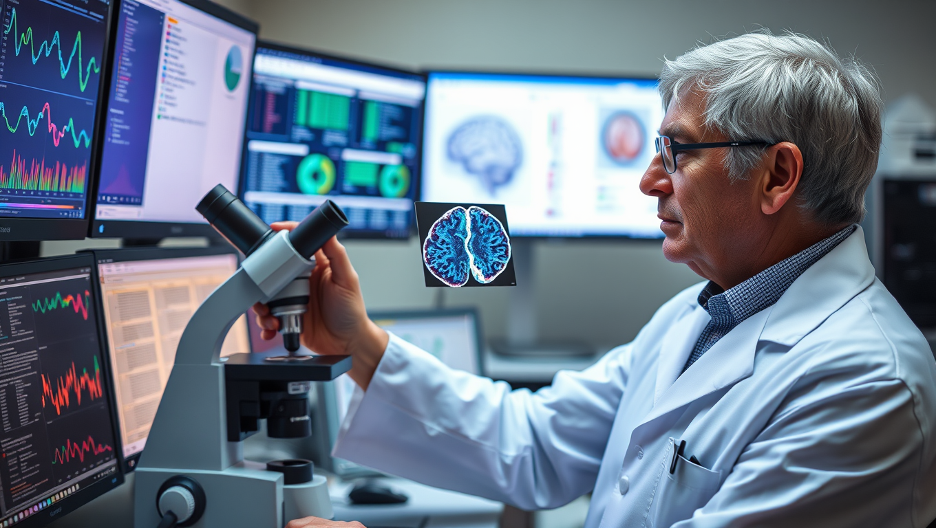
Reversing Alzheimer’s Damage: A Surprising Breakthrough with Cancer Drugs
In an exciting breakthrough, researchers have identified cancer drugs that might reverse the effects of Alzheimer’s disease in the brain. By analyzing gene expression in brain cells, they discovered that some FDA-approved cancer medications could reverse damage caused by Alzheimer’s.
Breast Cancer
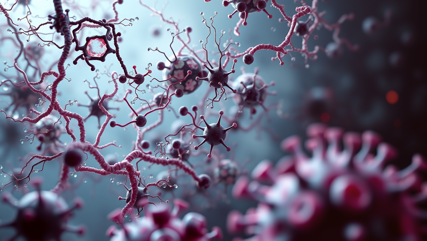
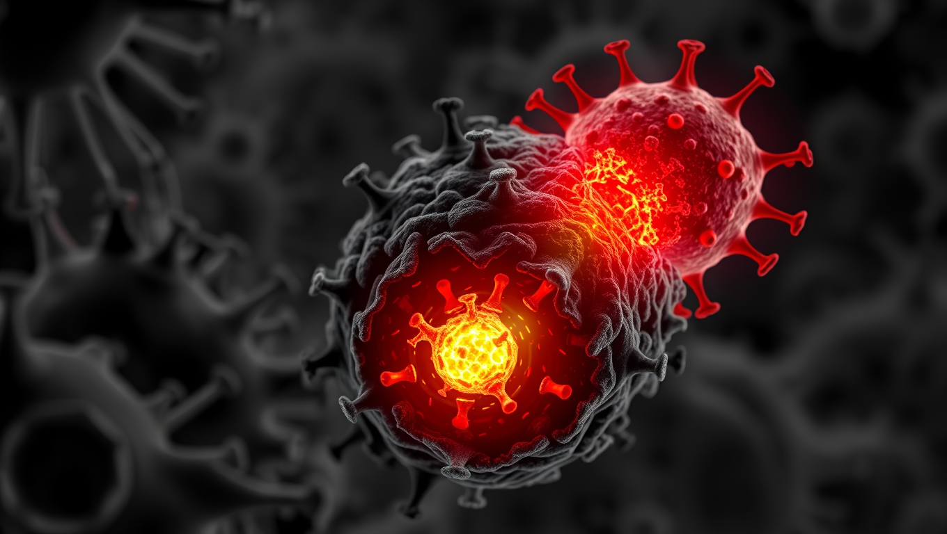
The Fatal Mutation That Lets Cancer Outsmart Our Immune System
Scientists at UC Davis discovered a small genetic difference that could explain why humans are more prone to certain cancers than our primate cousins. The change affects a protein used by immune cells to kill tumors—except in humans, it’s vulnerable to being shut down by an enzyme that tumors release. This flaw may be one reason treatments like CAR-T don’t work as well on solid tumors. The surprising twist? That mutation might have helped our brains grow larger over time. Now, researchers are exploring ways to block the enzyme and give our immune system its power back.
Anxiety
Single Psilocybin Dose Delivers Long-Term Depression Relief for Cancer Patients
Psilocybin, the active ingredient in magic mushrooms, might just revolutionize how depression and anxiety are treated in cancer patients. In a groundbreaking trial, a single dose combined with therapy significantly reduced emotional suffering, and these effects often lasted over two years. As follow-up studies expand the research to multiple doses and larger samples, scientists are eyeing a possible new standard of care that merges psychedelics with psychological support.


A New Horizon for Vision: How Gold Nanoparticles May Restore People’s Sight

Retiring Abroad Can Be Lonely Business

Revolutionizing Quantum Communication: Direct Connections Between Multiple Processors
Harnessing Water Waves: A Breakthrough in Controlling Floating Objects

“Unveiling Hidden Patterns: A New Twist on Interference Phenomena”

Household Electricity Three Times More Expensive Than Upcoming ‘Eco-Friendly’ Aviation E-Fuels, Study Reveals
Reducing Falls Among Elderly Women with Polypharmacy through Exercise Intervention
-
 Detectors11 months ago
A New Horizon for Vision: How Gold Nanoparticles May Restore People’s Sight
-
Earth & Climate12 months ago
Retiring Abroad Can Be Lonely Business
-
Cancer12 months ago
Revolutionizing Quantum Communication: Direct Connections Between Multiple Processors
-
Albert Einstein12 months ago
Harnessing Water Waves: A Breakthrough in Controlling Floating Objects
-
Chemistry11 months ago
“Unveiling Hidden Patterns: A New Twist on Interference Phenomena”
-
Earth & Climate12 months ago
Household Electricity Three Times More Expensive Than Upcoming ‘Eco-Friendly’ Aviation E-Fuels, Study Reveals
-
Diseases and Conditions12 months ago
Reducing Falls Among Elderly Women with Polypharmacy through Exercise Intervention
-
Agriculture and Food12 months ago
“A Sustainable Solution: Researchers Create Hybrid Cheese with 25% Pea Protein”


