Connect with us
We’re experimenting with AI-generated content to help deliver information faster and more efficiently.
While we try to keep things accurate, this content is part of an ongoing experiment and may not always be reliable.
Please double-check important details — we’re not responsible for how the information is used.

Biology

Unlocking Regeneration: Scientists Discover Key Factor in Axolotls’ Ability to Grow Limbs

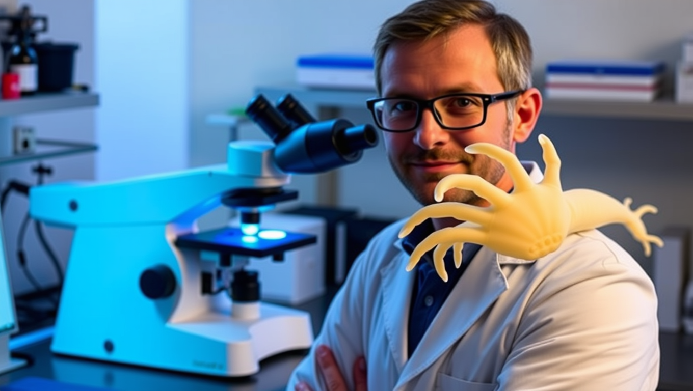
With its fascinating ability to regrow entire limbs and internal organs, the Mexican axolotl is the ideal model for studying regeneration. Scientists have now found a factor that tells cells which part of the arm to regenerate — and used it to reprogram the identity of cells as they develop. This breakthrough for the regeneration research field has implications for tissue engineering, including in human tissues.
Avatar photo

Published

on

The Mexican axolotl has long fascinated scientists with its incredible ability to regrow entire limbs and internal organs. A breakthrough study by Elly Tanaka’s lab at IMBA has now found a crucial factor that tells cells which part of the arm to regenerate – Hand2, a positional code that enables this remarkable feat. This discovery opens up promising possibilities for tissue engineering and regenerative therapies.

In a murky lake near Mexico City, axolotls live in constant danger of losing limbs to aggressive neighbors. However, their bodies have an extraordinary ability to regenerate lost limbs within as few as eight weeks. To achieve this, the regrowing body parts must “know” their position within the axolotl’s body to form the right structure for a specific location. The researchers found that cells on the anterior (thumb) side of the limb express FGF8, while posterior (pinky) side stem cells express Shh. These two signals reinforce each other to instruct cells to grow and pattern the regenerating arm.

A key breakthrough came when scientists discovered hundreds of factors that differed in the anterior versus the posterior half of the limb even before an injury. Hand2 was found to be expressed on the posterior side, acting as a positional cue. Experiments confirmed its crucial role in switching on Shh after injury, demonstrating Hand2’s central position in providing positional information.

The scientists developed a new model for limb regeneration, likening it to a radio broadcast: cells on the posterior side express Hand2 at low levels, maintaining their stable memory of being in the pinky zone. Upon injury, these cells dial up Hand2 expression to switch on Shh signals in a subset of Hand2-expressing cells. Cells near the Shh source regenerate as posterior-part-cells, while cells far away from the Shh signal regenerate as anterior-part-cells.

This finding can explain how preexisting positional memory signals reactivate upon injury to induce correct patterning. The discovery holds immense potential for tissue engineering and regenerative therapies, allowing scientists to “reprogram” cells to change their function.

The ability to alter cell identities is particularly promising, as it could enable scientists to transform cells in different parts of the body. Harnessing such signals might allow us to push cells beyond their normal biological limits, unlocking new roles for medical innovation.

The discovery that axolotls rely on the Hand2-Shh signaling circuit for limb regeneration is exciting, as these same genes are present in humans. This suggests that scientists may one day be able to target similar memory in human limbs to unlock new regenerative capabilities.

Animal Learning and Intelligence

The Buzz on Caffeine: How it Affects the Sleeping Brain

Coffee can help you stay awake. But what does caffeine actually do to your brain once you’re asleep? Using AI, a team of researchers has an answer: it affects the brain’s ‘criticality’.

Avatar photo

Published

on

The article delves into the intricate relationship between caffeine and the sleeping brain, offering fresh insights from a recent study published in Nature Communications Biology. Researchers from Université de Montréal have shed new light on how caffeine can modify sleep patterns and influence the brain’s recovery during the night.

Led by Philipp Thölke, a research trainee at UdeM’s Cognitive and Computational Neuroscience Laboratory (CoCo Lab), the team used AI and electroencephalography (EEG) to study caffeine’s effects on sleep. Their findings reveal that caffeine increases the complexity of brain signals and enhances brain “criticality” during sleep – a state characterized by balanced order and chaos.

Interestingly, this effect is more pronounced in younger adults, particularly during REM sleep, the phase associated with dreaming. The researchers attribute this finding to a higher density of adenosine receptors in young brains, which naturally decrease with age. Adenosine is a molecule that accumulates throughout the day, causing fatigue.

The study’s lead author, Thölke, notes that caffeine stimulates the brain and pushes it into a state of criticality, where it is more awake, alert, and reactive. However, this state can interfere with rest at night, preventing the brain from relaxing or recovering properly.

The researchers used EEG to record the nocturnal brain activity of 40 healthy adults on two separate nights: one when they consumed caffeine capsules three hours before bedtime and another when they took a placebo at the same time. They applied advanced statistical analysis and artificial intelligence to identify subtle changes in neuronal activity, revealing that caffeine increased the complexity of brain signals during sleep.

The team also discovered striking changes in the brain’s electrical rhythms during sleep: caffeine attenuated slower oscillations such as theta and alpha waves – generally associated with deep, restorative sleep – and stimulated beta wave activity, which is more common during wakefulness and mental engagement.

These findings suggest that even during sleep, the brain remains in a more activated, less restorative state under the influence of caffeine. This change in the brain’s rhythmic activity may help explain why caffeine affects the efficiency with which the brain recovers during the night, with potential consequences for memory processing.

The study’s implications are significant, particularly given the widespread use of caffeine as a daily remedy for fatigue. The researchers stress the importance of understanding its complex effects on brain activity across different age groups and health conditions. They add that further research is needed to clarify how these neural changes affect cognitive health and daily functioning, potentially guiding personalized recommendations for caffeine intake.

Continue Reading

Biology

Unraveling Microtubule Mysteries: Scientists Crack Code on Cellular Scaffolding Secrets

Scientists found out how naturally unstable filaments decide whether to grow or to shorten.

Avatar photo

Published

on

A groundbreaking study has shed new light on the fundamental mechanisms governing microtubule growth within cells. Researchers from Queen Mary University of London and the University of Dundee have made a significant breakthrough by discovering that the ability of tubulin proteins at microtubule ends to connect with each other sideways determines whether a microtubule elongates or shortens.

Microtubules are crucial protein structures that form the internal skeleton of cells, providing structural support and generating dynamic forces that push and pull. These tiny filaments constantly assemble and disassemble by adding or removing tubulin building blocks at their ends. However, the precise rules dictating whether a microtubule grows or shrinks have long remained a mystery due to the complexity and miniature size of their ends.

The collaborative research team has cracked part of this code using advanced computer simulations coupled with innovative imaging techniques. This interdisciplinary approach has allowed them to address this complex biological question from a fresh perspective, bridging physics and biology.

Dr. Vladimir Volkov, co-lead author from Queen Mary University of London, explained the significance of their findings: “Understanding how microtubules grow and shorten is very important – this mechanism underlies division and motility of all our cells. Our results will inform future biomedical research, particularly in areas related to cell growth and cancer.”

Dr. Maxim Igaev, co-lead author from the University of Dundee, highlighted the power of their interdisciplinary approach: “Bridging physics and biology has allowed us to address this complex biological question from a fresh perspective. This synergy not only enriches both fields but also paves the way for discoveries that neither discipline could achieve in isolation.”

This exciting research deepens our understanding of fundamental cellular processes and opens potential new avenues for biomedical research, particularly in areas concerning cell proliferation and the development of treatments for diseases like cancer.

Continue Reading

Biology

Unlocking Plant Defenses: Scientists Discover New Way to Help Plants Fight Diseases

Laboratory could improve crop resilience In a discovery three decades in the making, scientists have acquired detailed knowledge about the internal structures and mode of regulation for a specialized protein and are proceeding to develop tools that can capitalize on its ability to help plants combat a wide range of diseases. The work, which exploits a natural process where plant cells die on purpose to help the host plant stay healthy, is expected to have wide applications in the agricultural sector, offering new ways to protect major food crops from a variety of devastating diseases, the scientists said.

Avatar photo

Published

on

The discovery of scientists at Rutgers and Brookhaven National Laboratory has shed light on a natural process where plant cells intentionally die to help their host stay healthy. This phenomenon, known as programmed cell death or cell suicide, is crucial for fighting diseases and responding to stress in plants.

A team led by Eric Lam at Rutgers University-New Brunswick and Qun Liu at Brookhaven National Laboratory reported that advanced crystallography and computer modeling techniques have enabled them to obtain a detailed understanding of metacaspase 9, a pivotal plant protease. This enzyme plays a central role in programmed cell death and has been linked to two major types of disease-causing agents for plants: biotrophs and necrotrophs.

The researchers found that strengthening metacaspase 9 may prevent biotrophic diseases, while jamming its function means the enzyme won’t assist necrotrophs in killing healthy cells. By creating “super-active variants” of the enzyme, they may provide novel resistance traits to a slew of important diseases, such as powdery mildew and rusts.

This breakthrough has significant implications for agriculture, as it could lead to safer and more effective treatments for crops around the world. The researchers have already started exploring ways to develop tools that can harness metacaspase 9’s biological functions to protect plants from devastating diseases.

The team’s work was funded by the U.S. Department of Energy’s Office of Science and the National Science Foundation, and they used Highly Automated Macromolecular Crystallography (AMX) and Frontier Microfocusing Macromolecular Crystallography (FMX) beamlines at NSLS-II, a DOE Office of Science user facility.

This discovery is a testament to the power of scientific collaboration and the potential for groundbreaking research to improve our understanding of the natural world. As scientists continue to unravel the mysteries of plant biology, we may uncover new ways to protect crops from diseases and promote sustainable agriculture practices that benefit both people and the environment.

Continue Reading

Trending