
While we try to keep things accurate, this content is part of an ongoing experiment and may not always be reliable.
Please double-check important details — we’re not responsible for how the information is used.
Extreme Survival
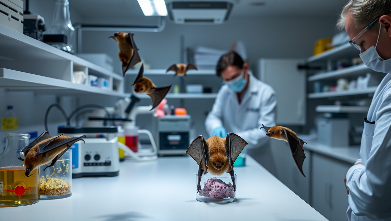
Unlocking the Secrets of Bat Immunity: A New Platform for Studying Viral Defense Mechanisms
Bats are known as natural hosts for highly pathogenic viruses such as MERS- and SARS-related coronaviruses, as well as the Marburg and Nipah viruses. In contrast to the severe and often fatal disease outcomes these viruses cause in humans, bats generally do not show obvious signs of viral illness following infection. An international research team has developed an innovative organoid research platform that allowed them to closely investigate the cellular antiviral defense mechanisms of mucosal epithelial tissues of bats. The results could pave the way for the development of new therapies against viral diseases.


Early Humans
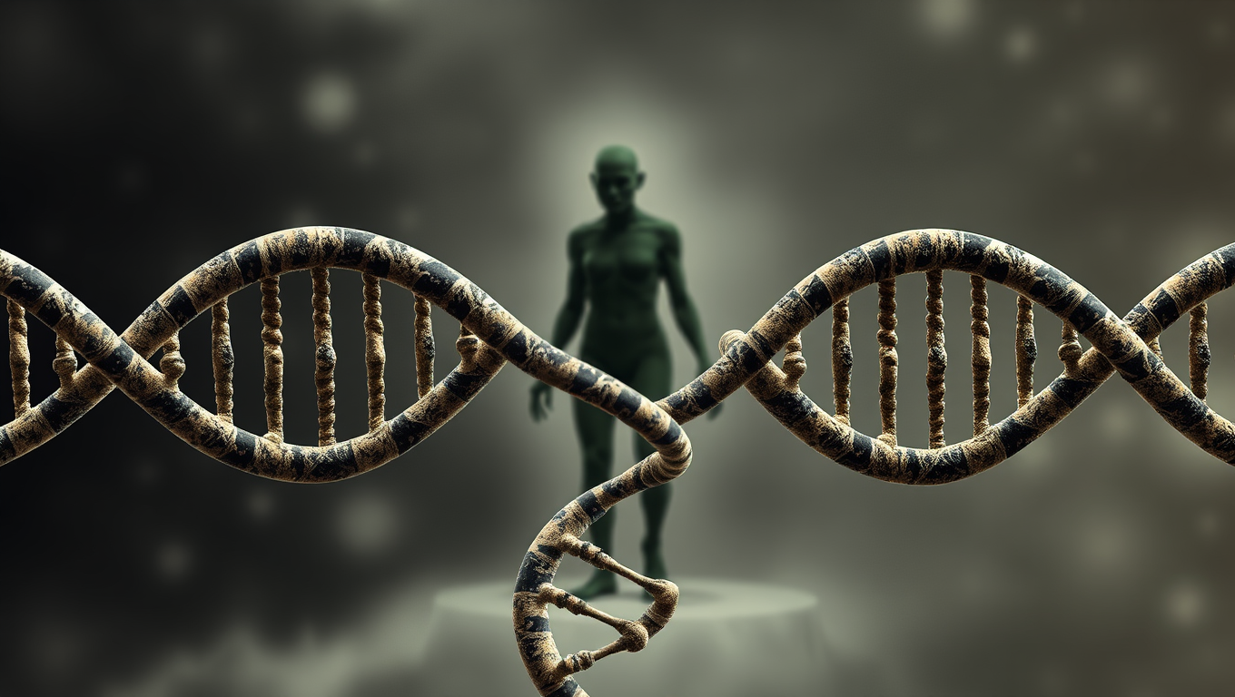
The Hidden Legacy of the Denisovans: Uncovering the Secrets of Human Evolution
Denisovans, a mysterious human relative, left behind far more than a handful of fossils—they left genetic fingerprints in modern humans across the globe. Multiple interbreeding events with distinct Denisovan populations helped shape traits like high-altitude survival in Tibetans, cold-weather adaptation in Inuits, and enhanced immunity. Their influence spanned from Siberia to South America, and scientists are now uncovering how these genetic gifts transformed human evolution, even with such limited physical remains.
Bacteria
Unlocking the Secrets of Mars: Cosmic Rays Reveal Hidden Potential for Life
Cosmic rays from deep space might be the secret energy source that allows life to exist underground on Mars and icy moons like Enceladus and Europa. New research reveals that when these rays interact with water or ice below the surface, they release energy-carrying electrons that could feed microscopic life, a process known as radiolysis. This breakthrough suggests that life doesn’t need sunlight or heat, just some buried water and radiation.

Agriculture and Food
The Ancient Origins of Potatoes Revealed
About 9 million years ago, a wild interspecies fling between tomato-like plants and potato relatives in South America gave rise to one of the world’s most important crops: the potato. Scientists have now traced its roots to a rare natural hybridization that created the tuber, a storage organ that allowed the plant to survive harsh Andean environments and spread rapidly.


A New Horizon for Vision: How Gold Nanoparticles May Restore People’s Sight

Retiring Abroad Can Be Lonely Business

Revolutionizing Quantum Communication: Direct Connections Between Multiple Processors
Harnessing Water Waves: A Breakthrough in Controlling Floating Objects

“Unveiling Hidden Patterns: A New Twist on Interference Phenomena”

Household Electricity Three Times More Expensive Than Upcoming ‘Eco-Friendly’ Aviation E-Fuels, Study Reveals
Reducing Falls Among Elderly Women with Polypharmacy through Exercise Intervention
-
 Detectors11 months ago
A New Horizon for Vision: How Gold Nanoparticles May Restore People’s Sight
-
Earth & Climate1 year ago
Retiring Abroad Can Be Lonely Business
-
Cancer12 months ago
Revolutionizing Quantum Communication: Direct Connections Between Multiple Processors
-
Albert Einstein1 year ago
Harnessing Water Waves: A Breakthrough in Controlling Floating Objects
-
Chemistry12 months ago
“Unveiling Hidden Patterns: A New Twist on Interference Phenomena”
-
Earth & Climate12 months ago
Household Electricity Three Times More Expensive Than Upcoming ‘Eco-Friendly’ Aviation E-Fuels, Study Reveals
-
Diseases and Conditions1 year ago
Reducing Falls Among Elderly Women with Polypharmacy through Exercise Intervention
-
Agriculture and Food12 months ago
“A Sustainable Solution: Researchers Create Hybrid Cheese with 25% Pea Protein”


