Connect with us
We’re experimenting with AI-generated content to help deliver information faster and more efficiently.
While we try to keep things accurate, this content is part of an ongoing experiment and may not always be reliable.
Please double-check important details — we’re not responsible for how the information is used.

COPD

Unlocking the Secrets of Deadly Lung Disease: Scientists Discover Immune Cell Networks Driving Idiopathic Pulmonary Fibrosis

Researchers reveal critical mechanism behind idiopathic pulmonary fibrosis.

Avatar photo

Published

on

A groundbreaking study published in the European Respiratory Journal has shed new light on the devastating lung disease Idiopathic Pulmonary Fibrosis (IPF). Researchers from Rutgers Health have identified networks of misplaced immune cells driving this aggressive condition, potentially opening a path to new treatments for IPF patients.

For decades, scientists have struggled to understand the underlying mechanisms of IPF, which scars lung tissue and makes breathing increasingly difficult until patients can’t get enough oxygen. The available drugs provide minimal benefit, and lung transplantation works for some patients, but transplants have a 50% five-year mortality rate.

The study used advanced spatial mapping techniques to compare healthy lung tissues and tissues from patients with fatal IPF. The researchers discovered that disease-scarred lung tissue abounds in plasma cells – specialized immune cells that typically reside in bone marrow and produce antibodies. This was a striking finding, as normal lungs have almost no plasma cells.

The researchers identified previously unknown cellular networks orchestrating this abnormal immune response. They discovered novel mural cells wrapping around blood vessels and producing signal proteins that organize immune responses. Unique fibroblasts secreting a protein that attracts plasma cells to damaged areas were also found.

This particular type of fibroblast has never been described before, and its role in scarring is still unknown. However, the researchers believe that targeting these abnormal cellular networks may prove an effective disease treatment in humans.

The research team began using live mice to see if reducing plasma in the lungs slowed disease formation. This work demonstrated that blocking signaling pathways reduced plasma cell accumulation and alleviated lung scarring. The findings suggest that drugs targeting plasma cells already exist, which could potentially be repurposed to treat IPF.

For patients with IPF, the findings offer hope of new treatments for a debilitating condition with limited therapeutic options. The disease typically affects men over 60 years of age, with most patients dying within five years of diagnosis.

The next steps for the research team include determining whether the plasma cells are producing autoantibodies against healthy tissues and further investigating how fibroblasts and mural cells develop their abnormal properties in IPF. This study represents a collaborative effort between the Child Health Institute of New Jersey and the Rutgers Institute for Translational Medicine and Science, combining mouse model research with analysis of human lung tissue from end-stage IPF patients.

“Our research suggests that IPF might have a strong autoimmune link,” said Qi Yang, an associate pediatrics professor at Rutgers Robert Wood Johnson Medical School.

COPD

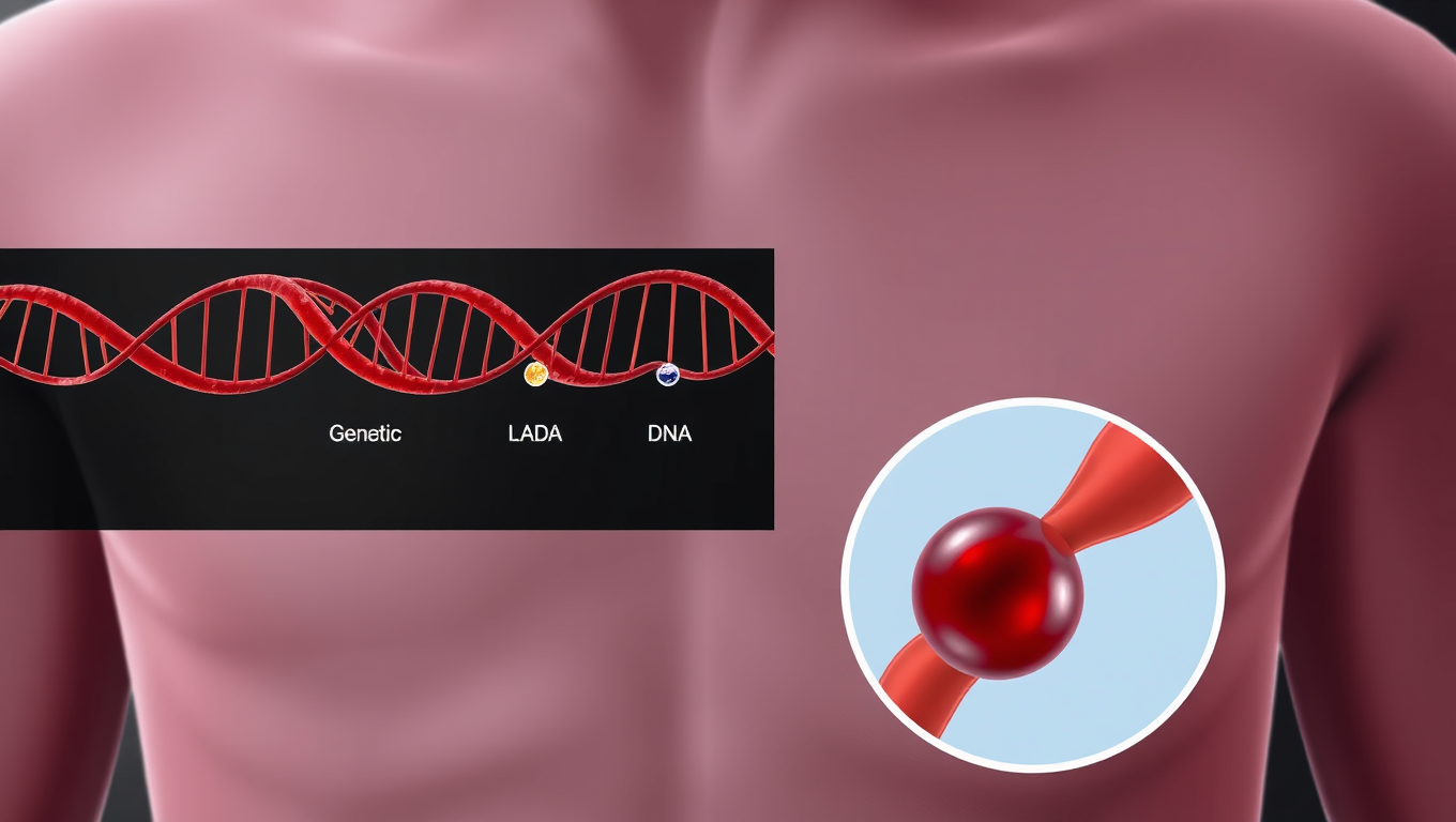
Revolutionary Nanoparticles Deliver Genetic Treatments Directly to the Lungs

A scientific team has unlocked a new way to treat serious lung conditions by using specially designed nanoparticles to deliver genetic therapies straight to lung cells. This innovation could transform care for patients with cystic fibrosis or lung cancer. With a powerful combination of gene editing and RNA delivery, the system has already shown promise in animal trials. The streamlined approach not only enhances precision but also avoids harmful side effects, making it a bold leap forward in respiratory medicine.

Avatar photo

Published

on

The fight against respiratory diseases has just received a significant boost. Scientists have developed a groundbreaking new drug delivery system that targets genetic therapies directly to the lungs. This innovative approach has opened promising possibilities for patients suffering from conditions like lung cancer and cystic fibrosis, offering a glimmer of hope for those in need.

Led by Gaurav Sahay from Oregon State University’s College of Pharmacy, this research was conducted in collaboration with Oregon Health & Science University and the University of Helsinki. The findings were published in two separate papers: one in Nature Communications and another in the Journal of the American Chemical Society.

Through an extensive study involving over 150 different materials, researchers discovered a novel type of nanoparticle that can safely and effectively carry messenger RNA and gene-editing tools to lung cells. In experiments with mice, this treatment demonstrated remarkable results – slowing down the growth of lung cancer and improving lung function that had been compromised by cystic fibrosis.

Cystic fibrosis is a genetic condition caused by one faulty gene, which severely limits lung function in patients. The new drug delivery system has shown great promise in addressing this issue, with researchers developing a chemical strategy to build a broad library of lung-targeting lipids used in the nanocarriers. These materials form the foundation for the innovative treatment and can be customized to reach different organs in the body.

“The streamlined synthesis method makes it easier to design future therapies for a wide range of diseases,” Sahay explained. “These results demonstrate the power of targeted delivery for genetic medicines. We were able to both activate the immune system to fight cancer and restore function in a genetic lung disease, without harmful side effects.”

The studies were funded by esteemed organizations such as the Cystic Fibrosis Foundation, the National Cancer Institute, and the National Heart, Lung and Blood Institute. This groundbreaking research has brought hope for patients with respiratory diseases, and its long-term implications could revolutionize the treatment of various conditions.

As Sahay put it, “Our long-term goal is to create safer, more effective treatments by delivering the right genetic tools to the right place. This is a major step in that direction.”

Continue Reading

Brain Tumor

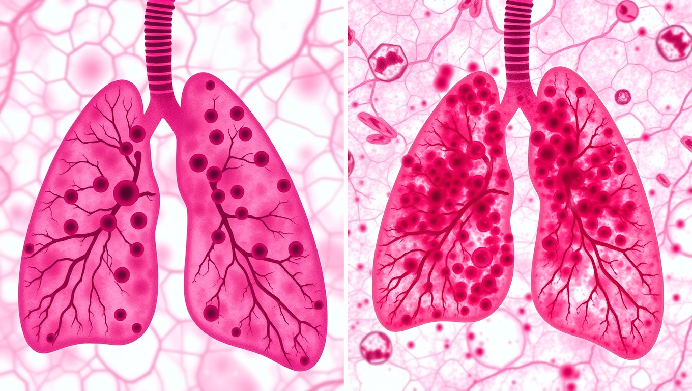
Uncovering the Secrets of COPD: The Role of Carbon Build-up in Lung Disease

Scientists have discovered that people with COPD have lung cells that contain over three times as much soot-like carbon as those of smokers without the disease. These overloaded cells are larger and trigger more inflammation, suggesting that pollution and carbon buildup not just smoking may drive the disease.

Avatar photo

Published

on

Chronic obstructive pulmonary disease (COPD) is a complex condition that affects millions worldwide. Researchers have long sought to understand the underlying causes of this debilitating disease. A recent study published in ERJ Open Research has shed new light on the role of carbon build-up in COPD, revealing a significant accumulation of soot-like deposits in the lungs of affected individuals.

The research team, led by Drs James Baker and Simon Lea from the University of Manchester, UK, investigated the impact of carbon exposure on alveolar macrophages – cells that protect the body by engulfing particles and bacteria. In COPD patients, these cells are found to be larger and more prone to inflammation when exposed to carbon.

The study compared samples from 28 people with COPD and 15 smokers without the disease. They discovered a staggering three-fold increase in carbon accumulation within alveolar macrophage cells of COPD patients compared to those who smoked but did not have COPD. Notably, patients with larger deposits of carbon in their alveolar macrophages exhibited worse lung function, as measured by FEV1%.

Dr Lea noted that this build-up of carbon is not a direct result of cigarette smoking, but rather an inherent difference in the form and function of alveolar macrophages between COPD patients and smokers. This raises intriguing questions about the causes of increased carbon levels in COPD patients’ macrophages. Is it because people with COPD are less able to clear the carbon they breathe in? Or is it due to exposure to more particulate matter, which accumulates and contributes to the development of COPD?

The implications of this research are significant, suggesting that reducing pollution in the air we breathe and helping people quit smoking may be crucial steps towards mitigating the risks of COPD. The study also highlights the need for further investigation into how carbon builds up over time and how lung cells respond.

As Professor Fabio Ricciardolo, Chair of the European Respiratory Society’s group on monitoring airway disease, pointed out, “This set of experiments suggests that people with COPD accumulate unusually large amounts of carbon in the cells of their lungs. This build-up seems to be altering those cells, potentially causing inflammation in the lungs and leading to worse lung function.”

The findings of this study offer valuable insights into the complex interplay between environmental and genetic factors in COPD. As researchers continue to unravel the mysteries of this disease, they hope to develop more effective treatments and preventive strategies for patients worldwide.

Continue Reading

Anemia

Hidden Risk: Three Genetic Variants That Raise Clot Risk by 180%

Genetic research in Sweden has unveiled three new gene variants that dramatically increase the risk of venous blood clots, sometimes by up to 180%. These discoveries build on existing knowledge of Factor V Leiden and suggest that genetics plays a bigger role than previously thought, especially for clots in the legs that can lead to life-threatening pulmonary embolisms.

Avatar photo

Published

on

The risk of blood clots is a serious concern worldwide, with venous thrombosis being one of the most common causes of death globally. A recent study from Lund University in Sweden has shed light on three genetic variants that significantly increase the risk of blood clots in the leg by up to 180%.

While arterial and venous blood clots have different causes and consequences, understanding the risk factors is crucial for prevention and treatment. In Sweden, over 10,000 people suffer from venous thromboembolism each year, with age being a strong risk factor.

“Venous thrombosis is a common disease that has always been somewhat overshadowed by arterial blood clots,” says Bengt Zöller, a specialist in general medicine at Skåne University Hospital and professor of general medicine at Lund University. “However, it’s essential to acknowledge its significance and take steps to prevent it.”
The risk factors for venous thrombosis include age, being overweight or tall, and lack of physical activity. Smoking is considered only a weak to moderate risk factor, while high blood pressure and high levels of blood lipids are associated with arterial clots, not venous ones.

Research suggests that commercial fishermen have a lower risk due to their diet rich in omega-3 fatty acids. Additionally, ultra-processed foods have been linked to an increased risk of blood clots, whereas plant-based diets may reduce this risk.
“Prophylaxis in the form of blood thinners may be particularly important if other risk factors are also present,” advises Zöller.

The three genetic variants identified by Bengt Zöller and his fellow researchers are ABO, F8, and VWF. These variants increase the risk of venous blood clots by 10-30% each, with an individual having five of these gene variants having a 180% higher risk.
“These genetic variants are present in all populations, making it essential to investigate how the number of risk genes affects the duration of treatment with anticoagulants after a blood clot,” concludes Zöller.

To prevent blood clots, one can take steps such as maintaining physical activity, monitoring blood pressure and lipid levels, quitting smoking, and eating a balanced diet rich in omega-3 fatty acids. Tailoring treatment based on risk assessment will become increasingly important in the future.
“Tailoring treatment based on risk assessment will become increasingly important,” concludes Bengt Zöller.

In summary, understanding the three genetic variants that increase the risk of blood clots by up to 180% is crucial for prevention and treatment. By acknowledging these risk factors, individuals can take steps to reduce their likelihood of developing venous thrombosis.
“Blood clot prevention is a vital aspect of healthcare, and awareness about the risks is essential,” emphasizes Zöller.

Continue Reading

Trending