Connect with us
We’re experimenting with AI-generated content to help deliver information faster and more efficiently.
While we try to keep things accurate, this content is part of an ongoing experiment and may not always be reliable.
Please double-check important details — we’re not responsible for how the information is used.

Diseases and Conditions

Unlocking the Secrets of Huntington’s Disease: Two Proteins with Opposing Effects

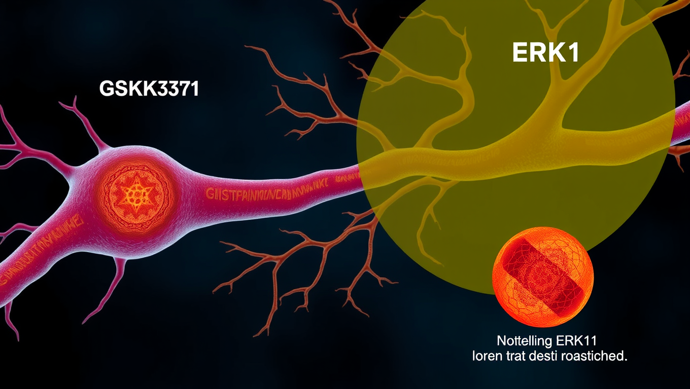
Researchers found that inhibiting GSK-3 led to less defects in the axonal transport process and less neuronal cell death, while inhibiting ERK1 led to more transport problems and more cell death.
Avatar photo

Published

on

The study of Huntington’s disease has been a longstanding mystery in neuroscience. Researchers at the University of Buffalo have made significant progress in understanding the disease by identifying two specific signaling proteins that play opposing roles in its progression.

The mutated huntingtin protein (HTT) is responsible for causing Huntington’s disease, but how it leads to the degeneration of neurons remains unclear. However, researchers have found that HTT functions as a traffic controller inside neurons, moving different cargo along axons with the help of other proteins. Reducing the amount of non-mutant HTT can lead to neurological problems.

In this study, the researchers focused on two signaling proteins: GSK3ß and ERK1. They were expressed more in the neurons of Huntington’s disease patients than in normal neurons. The team used fruit fly larvae with a mutant HTT to understand how these proteins affect neuronal function.

When they inhibited GSK3ß, they found that it led to less defects in axonal transport and reduced neuronal cell death. On the other hand, inhibiting ERK1 resulted in more axonal blockages and cell death.

The researchers suggest that ERK1 may protect neurons in the face of Huntington’s disease, while GSK3ß may exacerbate the condition. This means that therapeutics could potentially target these signaling proteins differently to treat this severe neurological disorder.

The study was supported by several organizations, including the National Institute of Neurological Disorders and Stroke, the Mark Diamond Research Fund, and the BrightFocus Foundation.

This research is a significant step towards understanding the progression of Huntington’s disease. By identifying two proteins with opposing effects, researchers can now explore ways to develop treatments that target these proteins differently. This could potentially lead to new therapies for this devastating disease.

Autism

Unpacking the Gene That Hijacks Fear: How PTEN Rewires the Brain’s Anxiety Circuit

Deleting a gene called PTEN in certain brain cells disrupts the brain’s fear circuitry and triggers anxiety-like behavior in mice — key traits seen in autism. Researchers mapped how this genetic tweak throws off the brain’s delicate balance of excitation and inhibition in the amygdala, offering deep insights into how one gene can drive specific ASD symptoms.

Avatar photo

Published

on

The gene PTEN has emerged as one of the most significant autism risk genes. Variations in this gene are found in a significant proportion of people with autism who also exhibit brain overgrowth. Researchers at the Max Planck Florida Institute for Neuroscience have discovered how loss of this gene rewires circuits and alters behavior, leading to increased fear learning and anxiety in mice – core traits seen in ASD.

PTEN has been linked to alterations in the function of inhibitory neurons in the development of ASD. The researchers focused on the changes in the central lateral amygdala driven by loss of PTEN in a critical neuronal population – somatostatin-expressing inhibitory neurons. They found that deleting PTEN specifically in these interneurons disrupted local inhibitory connectivity in the amygdala by roughly 50% and reduced the strength of the remaining inhibitory connections.

This diminished connectivity between inhibitory connections within the amygdala was contrasted by an increase in the strength of excitatory inputs received from the basolateral amygdala, a nearby brain region that relays emotionally-relevant sensory information to the amygdala. Behavioral analysis demonstrated that this imbalance in neural signaling was linked to heightened anxiety and increased fear learning, but not alterations in social behavior or repetitive behavior traits commonly observed in ASD.

The results confirm that PTEN loss in this specific cell type is sufficient to induce specific ASD-like behaviors and provide one of the most detailed maps to date of how local inhibitory networks in the amygdala are affected by genetic variations associated with neurological disorders. Importantly, the altered circuitry did not affect all ASD-relevant behaviors – social interactions remained largely intact – suggesting that PTEN-related anxiety and fear behaviors may stem from specific microcircuit changes.

By teasing out the local circuitry underlying specific traits, researchers hope to differentiate the roles of specific microcircuits within the umbrella of neurological disorders, which may one day help in developing targeted therapeutics for specific cognitive and behavioral characteristics. In future studies, they plan to evaluate these circuits in different genetic models to determine if these microcircuit alterations are convergent changes that underlie heightened fear and anxiety expression across diverse genetic profiles.

Continue Reading

Allergy

The Hidden Dangers of Fire Smoke Exposure

Smoke from wildfires and structural fires doesn t just irritate lungs it actually changes your immune system. Harvard scientists found that even healthy people exposed to smoke showed signs of immune system activation, genetic changes tied to allergies, and even toxic metals inside their immune cells.

Avatar photo

Published

on

The dangers of fire smoke exposure are well-documented, but until now, the full extent of its impact on our bodies has been unclear. A recent study led by researchers at Harvard T.H. Chan School of Public Health reveals that fire smoke can alter our immune system on a cellular level, leaving lasting changes and increasing our risk of serious health problems.

The study examined blood samples from 31 individuals who had been exposed to fire smoke and compared them to those from 29 non-exposed individuals. The results showed significant changes in the immune cells of those who had been exposed to smoke. These changes included an increase in memory CD8+ T cells, which are crucial for long-term immunity against pathogens, as well as elevated activation and chemokine receptor biomarkers that indicate inflammation and immune activity.

The researchers also found changes in 133 genes related to allergies and asthma in the individuals who had been exposed to smoke. Moreover, their immune cells were more likely to be bound with toxic metals like mercury and cadmium, which can further harm our health.

“This study fills a critical knowledge gap by showing exactly how fire smoke exposure can damage the body,” said Kari Nadeau, corresponding author of the study and chair of the Department of Environmental Health. “Our findings have significant implications for public health leaders and clinicians who need to respond to the growing threat of wildfires.”
The study’s lead author, Mary Johnson, added that the immune system is extremely sensitive to environmental exposures like fire smoke, even in healthy individuals. Knowing exactly how smoke exposure can harm our bodies may help us detect immune dysfunction earlier and pave the way for new therapeutics to mitigate or prevent the health effects of smoke exposure.

The researchers also noted that their study could inform environmental and public health policies and investments, such as increasing public awareness about the dangers of smoke exposure and the importance of following evacuation procedures during wildfires.
The study was funded by several organizations, including the National Institute of Environmental Health Sciences, the National Heart, Lung, and Blood Institute, and the San Francisco Cancer Prevention Foundation.

In conclusion, this study highlights the need for increased caution when it comes to fire smoke exposure. By understanding the full extent of its impact on our bodies, we can take steps to protect ourselves and others from its toxic effects.

Continue Reading

Allergy

The Resilient Enemy: Why Asthma Symptoms Persist Despite Powerful Drugs

Biological drugs have been a game-changer for people with severe asthma, helping them breathe easier and live more comfortably. But researchers at Karolinska Institutet have uncovered a surprising twist: while these treatments ease symptoms, they may not fully eliminate the immune cells that drive inflammation. In fact, some of these cells actually increase during treatment, suggesting the medication is managing symptoms without targeting the root cause. This could explain why asthma often returns when the drugs are stopped, raising questions about how long-term these treatments should be and whether we’re truly solving the underlying problem.

Avatar photo

Published

on

Asthma has long been a formidable foe for many people, causing symptoms that can range from mild discomfort to life-threatening attacks. While powerful biological drugs have significantly improved the lives of those with severe asthma, a recent study has shed light on why these symptoms often return despite treatment.

Biological drugs, or biologics, have become a crucial tool in managing severe asthma by helping patients keep their symptoms under control. However, researchers at Karolinska Institutet in Sweden discovered that certain immune cells, which play a significant role in asthma inflammation, do not disappear during treatment as previously thought. Instead, these inflammatory cells increase in number.

This finding suggests that biologics might not address the root cause of asthma, but rather manage its symptoms. As such, continued treatment may be necessary to keep the disease under control. This is particularly concerning considering that little is still known about the long-term effects of biologics like mepolizumab and dupilumab, which have been prescribed to asthmatics for less than ten years.

The study analyzed blood samples from 40 patients before and during treatment, using advanced methods such as flow cytometry and single-cell sequencing. Researchers were surprised to find that the levels of inflammatory cells in these patients increased rather than decreased. This could explain why inflammation of the airways often returns when the treatment is tapered or discontinued.

It is essential for researchers and medical professionals to understand the long-term immunological effects of biologics, as this knowledge can lead to more effective treatments and better outcomes for patients with severe asthma. The next stage of the study will involve analyzing samples from patients with a long treatment history and studying lung tissue to see how immune cells are affected in the airways.

The findings of this study have significant implications for the management and treatment of asthma, highlighting the need for continued research into the effects of biologics on the immune system.

Continue Reading

Trending