Connect with us
We’re experimenting with AI-generated content to help deliver information faster and more efficiently.
While we try to keep things accurate, this content is part of an ongoing experiment and may not always be reliable.
Please double-check important details — we’re not responsible for how the information is used.

Biology

Unlocking the Secrets of mRNA Vaccines: A Breakthrough in Cellular Regulation Revealed

A team of researchers has uncovered a key cellular mechanism that affects the function of mRNA vaccines and therapeutics. Their study provides the first comprehensive understanding of how mRNA vaccines are delivered, processed, and degraded within cells — a breakthrough that could pave the way for more effective vaccines and RNA-based treatments.
Avatar photo

Published

on

A team of researchers led by Dr. KIM V. Narry has made a groundbreaking discovery in understanding how mRNA vaccines are delivered, processed, and degraded within cells. Their study, published in Science, sheds light on the cellular mechanisms that affect the function of mRNA vaccines and therapeutics, paving the way for more effective treatments.

Messenger RNA (mRNA) plays a crucial role in mRNA vaccines, such as those used for COVID-19, and is also a promising tool for treating diseases like cancer and genetic disorders. When foreign mRNA enters cells, it must evade the body’s natural defense mechanisms to be effective. However, the detailed mechanisms by which mRNA is regulated inside cells have remained largely unknown.

The research team employed CRISPR-based knockout screening to identify the cellular factors involved in the delivery of mRNA into cells. This approach revealed three key factors that facilitate the cellular uptake or surveillance of exogenous mRNAs:

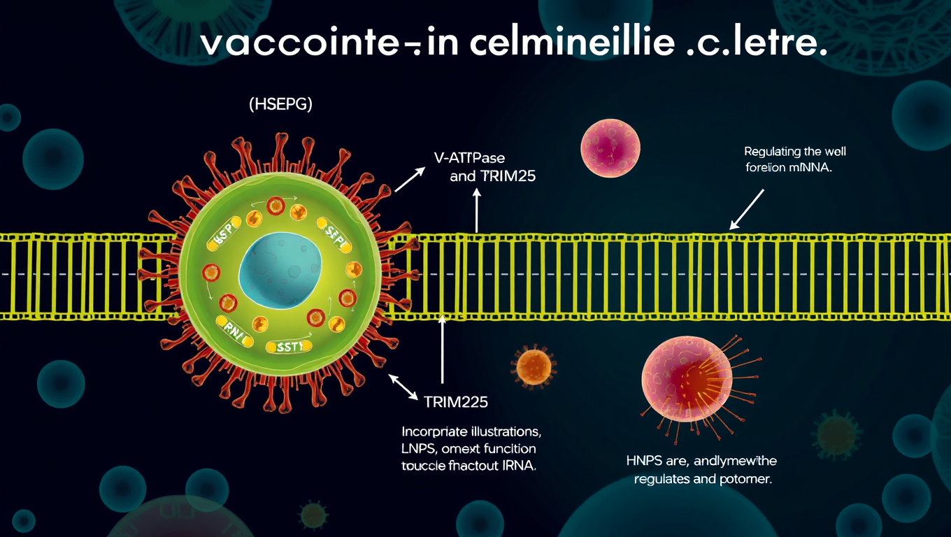
1. Heparan sulfate (HSPG), a sulfated glycoprotein on the cell surface, plays a crucial role in attracting LNPs and facilitating mRNA entry into the cell.
2. V-ATPase, a proton pump at the endosome, acidifies the vesicle and causes LNPs to become positively charged, enabling them to temporarily disrupt the endosomal membrane and release the mRNA into the cytoplasm.
3. TRIM25, a protein involved in the cellular defense mechanism, binds to and induces the rapid degradation of exogenous mRNAs, preventing their function.

The study highlights that mRNA molecules containing a special modification called N1-methylpseudouridine (m1Ψ) can evade TRIM25 detection, enhancing the stability and effectiveness of mRNA vaccines. This discovery emphasizes the importance of this modification in enhancing the therapeutic potential of mRNA-based treatments.

Additionally, the research demonstrates that proton ions serve as immune signaling molecules, providing new insights into how cells protect themselves from foreign RNA.

Dr. KIM V. Narry emphasized the importance of understanding these processes, stating, “Understanding how cells respond to mRNA vaccines is key to improving mRNA therapeutics. To develop effective RNA treatments, we need to find ways to bypass the cellular defense mechanisms and harness the endosomal system effectively.”

This research paves the way for more efficient mRNA vaccine delivery and offers a framework for future development of RNA-based therapies. The findings underscore the critical importance of early intervention and provide new directions for developing more effective treatments for a variety of diseases.

Behavioral Science

Echidna Pseudo-Pouch Microbiome Shifts During Lactation Helps Young Thrive

Research shows microbial communities in echidna pseudo-pouches undergo dramatic changes while the animal is lactating, which could help in creating an environment for their young, known as puggles, to thrive.

Avatar photo

Published

on

Echidnas, the only mammals that lay eggs, have an unusual reproductive system that includes a pseudo-pouch where their young, called puggles, grow and develop during lactation. Researchers from the University of Adelaide have made a fascinating discovery about the microbiome in these pseudo-pouches, which changes significantly while the mother is nursing her young.

The study, published in FEMS Microbiology Ecology, reveals that the microbial communities in echidna pseudo-pouches undergo dramatic changes during lactation, creating an environment that’s conducive to the health and well-being of their puggles. This is particularly important since puggles hatch at a very early developmental stage, lacking a functional immune system.

“We know that the reproductive microbiome is crucial for infant health in many species, including humans,” says Isabella Wilson, lead researcher on the study. “However, little was known about how it functions in egg-laying monotremes like echidnas.”

One of the key findings of this research is that during lactation, the pseudo-pouch microbial communities show significant differences in composition compared to samples taken outside of breeding season or during courtship and mating. This suggests that the echidna pseudo-pouch environment changes during lactation to accommodate young that lack a functional adaptive immune system.

The way puggles suckle may contribute to this shift in microbes. Unlike other species, echidnas don’t have nipples; instead, their young rub their beaks against a part of the pseudo-pouch called the milk patch, causing milk to come out of the skin, similar to a sweat or oil gland.

Compounds within the milk and from the skin probably contribute to the changes seen in the pseudo-pouch microbiota during lactation. This study highlights the importance of understanding these unique reproductive dynamics for conservation efforts and breeding programs for echidnas.

The research also sheds light on previous findings that showed big differences in the gut microbiome between echidnas in zoos and those in the wild. Surprisingly, no major difference was found in the pseudo-pouch microbiota between zoo-managed and wild animals. This suggests that the milk, rather than external environmental factors like captivity, is what primarily shapes the bacterial landscape of the pseudo-pouch.

For conservation efforts and breeding programs, it’s essential to learn more about the bacteria found in echidna pseudo-pouches and how they affect echidna health. This knowledge will help ensure the well-being of these unique animals and their young, ultimately contributing to the preservation of this fascinating species.

Continue Reading

Biochemistry Research

A More Realistic Look at DNA in Action: Researchers Shed Light on the Mechanical Forces Behind Strand Separation

By creating a more true-to-life representation of DNA’s environment, researchers have discovered that strand separation may take more mechanical force than the field previously believed.

Avatar photo

Published

on

The way we study DNA has long been a topic of discussion among researchers. Traditionally, biochemistry labs isolate DNA within a water-based solution that allows scientists to manipulate it without interacting with other molecules. However, this approach can be misleading, as it doesn’t reflect the true environment of a living cell. In fact, the interior of a cell is “super crowded” with molecules, which can significantly impact the behavior of DNA.

Researchers at Northwestern University have taken a more realistic approach to studying DNA by creating an environment that mimics the conditions within a living cell. Led by Professor John Marko, the team used microscopic magnetic tweezers to separate DNA and then carefully attach strands of it to surfaces on one end, and tiny magnetic particles on the other. This allowed them to conduct high-tech imaging and investigate how different types of molecules interact with DNA.

The researchers found that strand separation, a crucial process for initiating replication or making repairs, may require more mechanical force than previously believed. They introduced three types of molecules to the solution holding DNA, mimicking proteins and investigating interactions among glycerol, ethylene glycol, and polyethylene glycol (each approximately the size of one DNA double helix, two or three nanometers).

“We wanted to have a wide variety of molecules where some cause dehydration, destabilizing DNA mechanically, and then others that stabilize DNA,” said Northwestern post-doctoral researcher Parth Desai. “It’s not exactly analogous to things found in cells, but you could imagine that other competing proteins in cells will have a similar effect.”
The team wrote a paper on their findings, which will be published on June 17 in the Biophysical Journal. Marko and Desai hope to run more experiments that incorporate multiple crowding agents and move closer to a true representation of a cell.

“If this affects DNA strand separation, all protein interactions with DNA are also going to be affected,” said Marko. “For example, the tendency for proteins to stick to specific sites on DNA and to control specific processes — this is also going to be altered by crowding.”

Their research has significant implications for understanding fundamental biochemical processes and may lead to new medical advances. The team hopes to study how interactions between enzymes and DNA are impacted by crowding in a living cell, which could have far-reaching consequences for our understanding of cellular biology.

This work was supported by the National Institutes of Health (grant R01-GM105847) and by subcontract to the University of Massachusetts Center for 3D Structure and Physics of the Genome (under NIH grant UM1-HG011536).

Continue Reading

Animals

A “Roadmap” to Understanding the Fruit Fly Brain: Breakthrough Study Reveals Comprehensive Insights into Entire Nervous System

Researchers have gained comprehensive insights into the entire nervous system of the fruit fly (Drosophila melanogaster). The study describes in detail the neurons that span the entire nervous system of the adult fruit fly. The researchers also compared the complete set of neural connections (the connectome) in a female and a male specimen — and identified differences.

Avatar photo

Published

on

The fruit fly (Drosophila melanogaster) has long been a model organism for scientists studying genetics, development, and behavior. However, despite its importance, the intricacies of the fruit fly’s nervous system have remained somewhat of a mystery – until now. Researchers at Leipzig University and other institutions have made a groundbreaking discovery, publishing a study in Nature that provides comprehensive insights into the entire nervous system of the adult fruit fly.

For the first time, scientists have mapped out the neural connections (the connectome) in a female and a male specimen, revealing differences between the two sexes. This breakthrough is a significant step forward in understanding the complex interactions within the fruit fly’s brain and nervous system.

The study, led by Dr. Katharina Eichler from Leipzig University, involved analyzing three connectomes: one female brain data set and two nerve cord data sets (one male, one female). The researchers used light microscopy to identify all neurons in the neck of the fruit fly that could be visualized using this technique.

This allowed them to analyze the circuits formed by these cells in their entirety. When comparing male and female neurons, the scientists identified sex-specific differences for the first time. They found previously unknown cells that exist only in one sex and are absent in the other.

One notable example is a descending neuron known as aSP22, which communicates with neurons present only in females. This finding provides an explanation for the behavioral differences observed when this neuron is active: female flies extend their abdomen to lay eggs, while males curl theirs forward to mate.

The study’s findings are significant not only because they provide a comprehensive overview of the fruit fly connectome but also because they offer a “roadmap” for future research. By understanding the intricate connections within the nervous system, scientists can design more intelligent experiments to investigate the function of individual neurons or entire circuits – saving time and resources.

As Eichler notes, now that the technical challenges in analyzing the fruit fly’s nervous system have been overcome, her research group is working on two new data sets covering the entire central nervous system of both a female and a male specimen. This continued research will undoubtedly shed more light on the complexities of the fruit fly brain and its implications for our understanding of nervous systems in general.

Continue Reading

Trending