
While we try to keep things accurate, this content is part of an ongoing experiment and may not always be reliable.
Please double-check important details — we’re not responsible for how the information is used.
Brain Injury
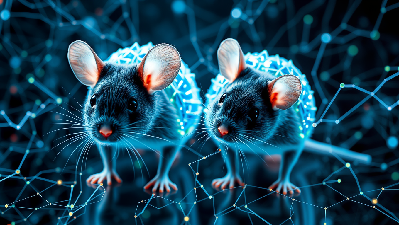
Unlocking the Secrets of the Brain with Digital Twins
In a new study, researchers created an AI model of the mouse visual cortex that predicts neuronal responses to visual images.


Alzheimer's
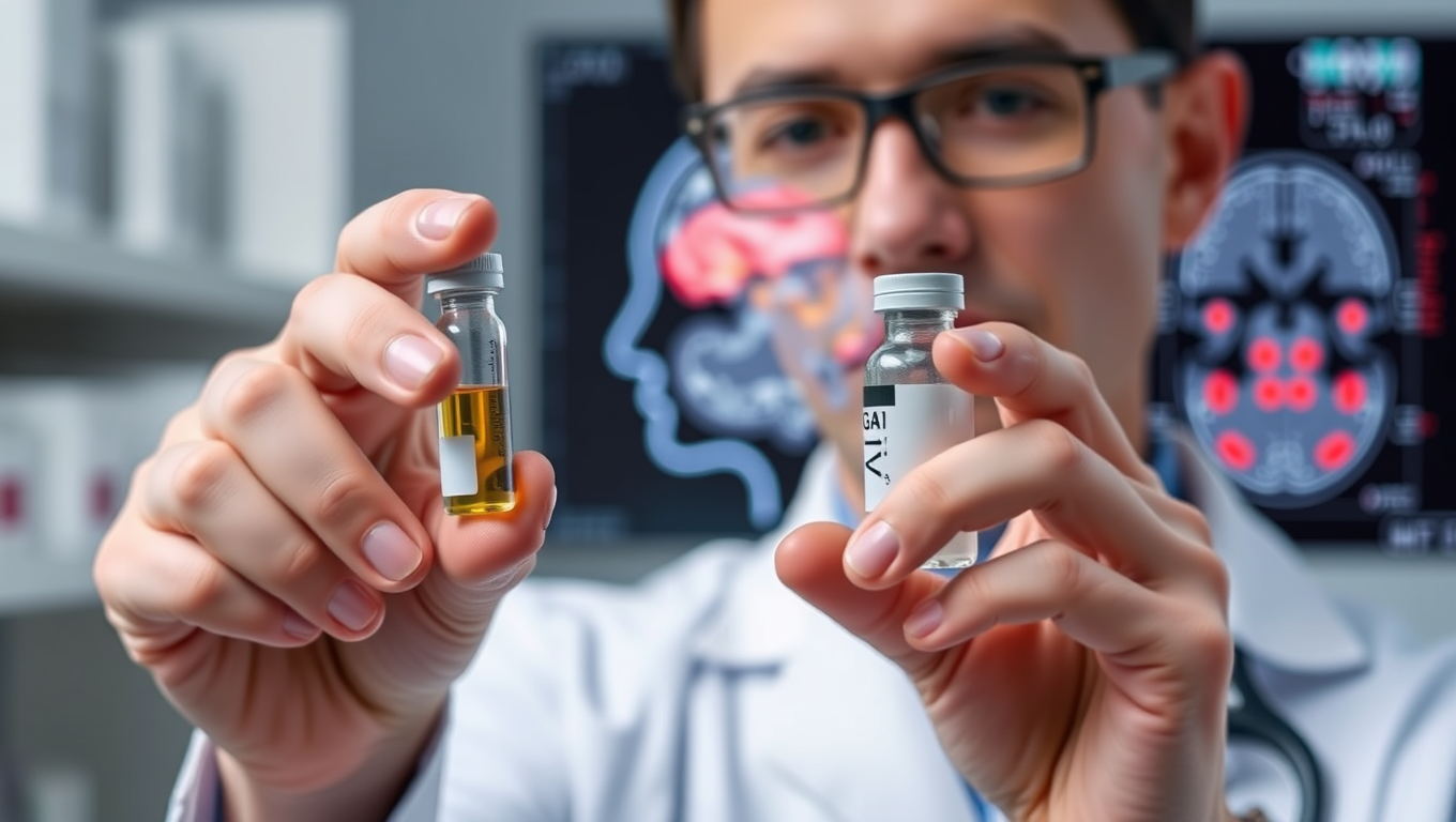
Rewinding Stroke Damage and Beyond: The Promise of GAI-17
Stroke kills millions, but Osaka researchers have unveiled GAI-17, a drug that halts toxic GAPDH clumping, slashes brain damage and paralysis in mice—even when given six hours post-stroke—and shows no major side effects, hinting at a single therapy that could also tackle Alzheimer’s and other tough neurological disorders.
Brain Injury
Scientists Edge Closer to Reversing Parkinson’s Symptoms — A Breakthrough for Humans?
Scientists at the University of Sydney have uncovered a malfunctioning version of the SOD1 protein that clumps inside brain cells and fuels Parkinson’s disease. In mouse models, restoring the protein’s function with a targeted copper supplement dramatically rescued movement, hinting at a future therapy that could slow or halt the disease in people.

Alzheimer's
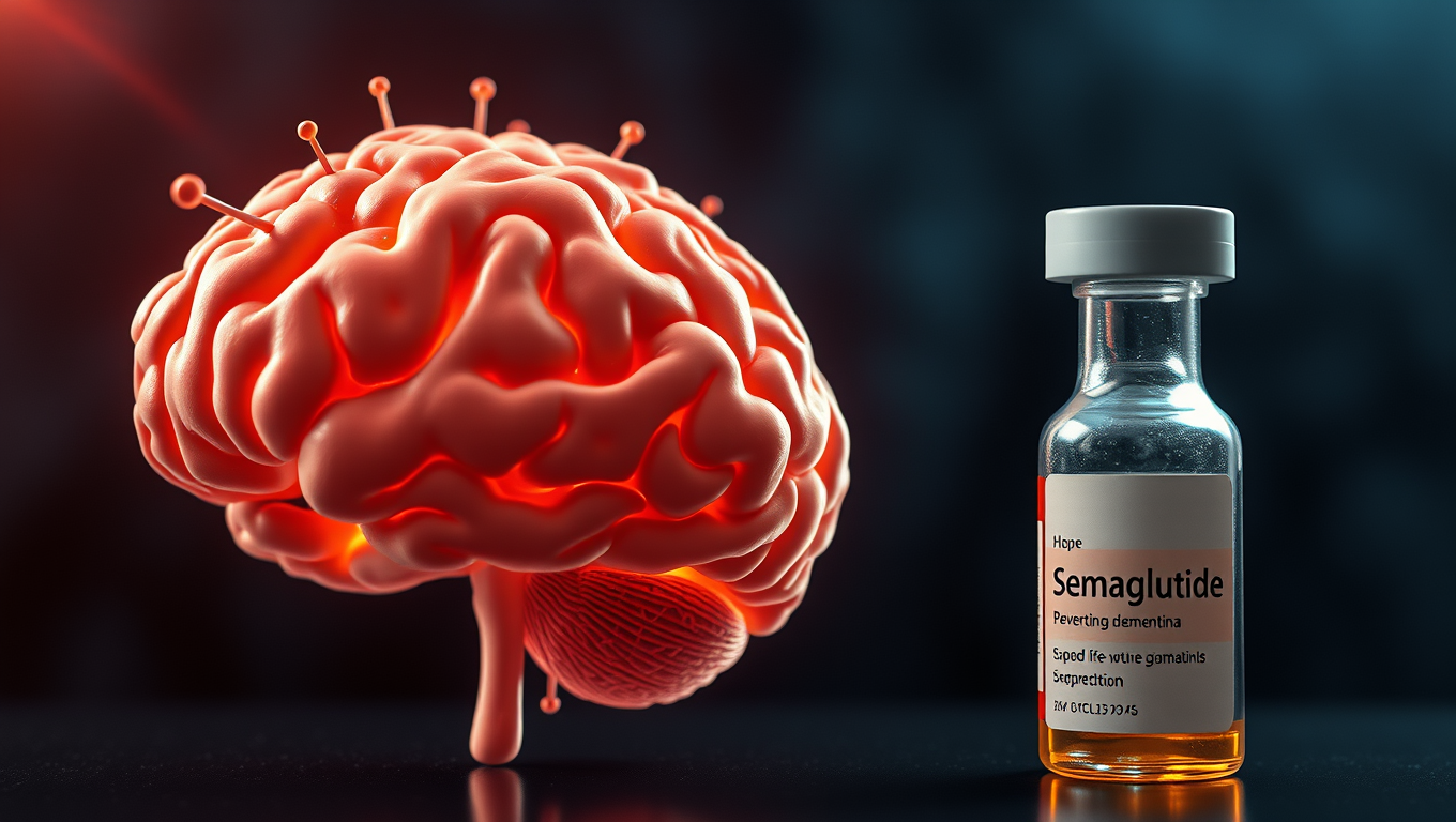
Groundbreaking Study Suggests Link Between Semaglutide and Lower Dementia Risk in Type 2 Diabetes Patients
A blockbuster diabetes and weight-loss drug might be doing more than controlling blood sugar—it could also be protecting the brain. Researchers at Case Western Reserve University found that people with type 2 diabetes who took semaglutide (the active ingredient in Ozempic and Wegovy) had a significantly lower risk of developing dementia. The benefit was especially strong in women and older adults.

A New Horizon for Vision: How Gold Nanoparticles May Restore People’s Sight

Retiring Abroad Can Be Lonely Business

Revolutionizing Quantum Communication: Direct Connections Between Multiple Processors
Harnessing Water Waves: A Breakthrough in Controlling Floating Objects

“Unveiling Hidden Patterns: A New Twist on Interference Phenomena”

Household Electricity Three Times More Expensive Than Upcoming ‘Eco-Friendly’ Aviation E-Fuels, Study Reveals
Reducing Falls Among Elderly Women with Polypharmacy through Exercise Intervention
-
 Detectors12 months ago
A New Horizon for Vision: How Gold Nanoparticles May Restore People’s Sight
-
Earth & Climate1 year ago
Retiring Abroad Can Be Lonely Business
-
Cancer1 year ago
Revolutionizing Quantum Communication: Direct Connections Between Multiple Processors
-
Albert Einstein1 year ago
Harnessing Water Waves: A Breakthrough in Controlling Floating Objects
-
Chemistry1 year ago
“Unveiling Hidden Patterns: A New Twist on Interference Phenomena”
-
Earth & Climate1 year ago
Household Electricity Three Times More Expensive Than Upcoming ‘Eco-Friendly’ Aviation E-Fuels, Study Reveals
-
Diseases and Conditions1 year ago
Reducing Falls Among Elderly Women with Polypharmacy through Exercise Intervention
-
Agriculture and Food1 year ago
“A Sustainable Solution: Researchers Create Hybrid Cheese with 25% Pea Protein”


