Connect with us
We’re experimenting with AI-generated content to help deliver information faster and more efficiently.
While we try to keep things accurate, this content is part of an ongoing experiment and may not always be reliable.
Please double-check important details — we’re not responsible for how the information is used.

Brain Injury

Unveiling the Hidden Brain Network for Naming

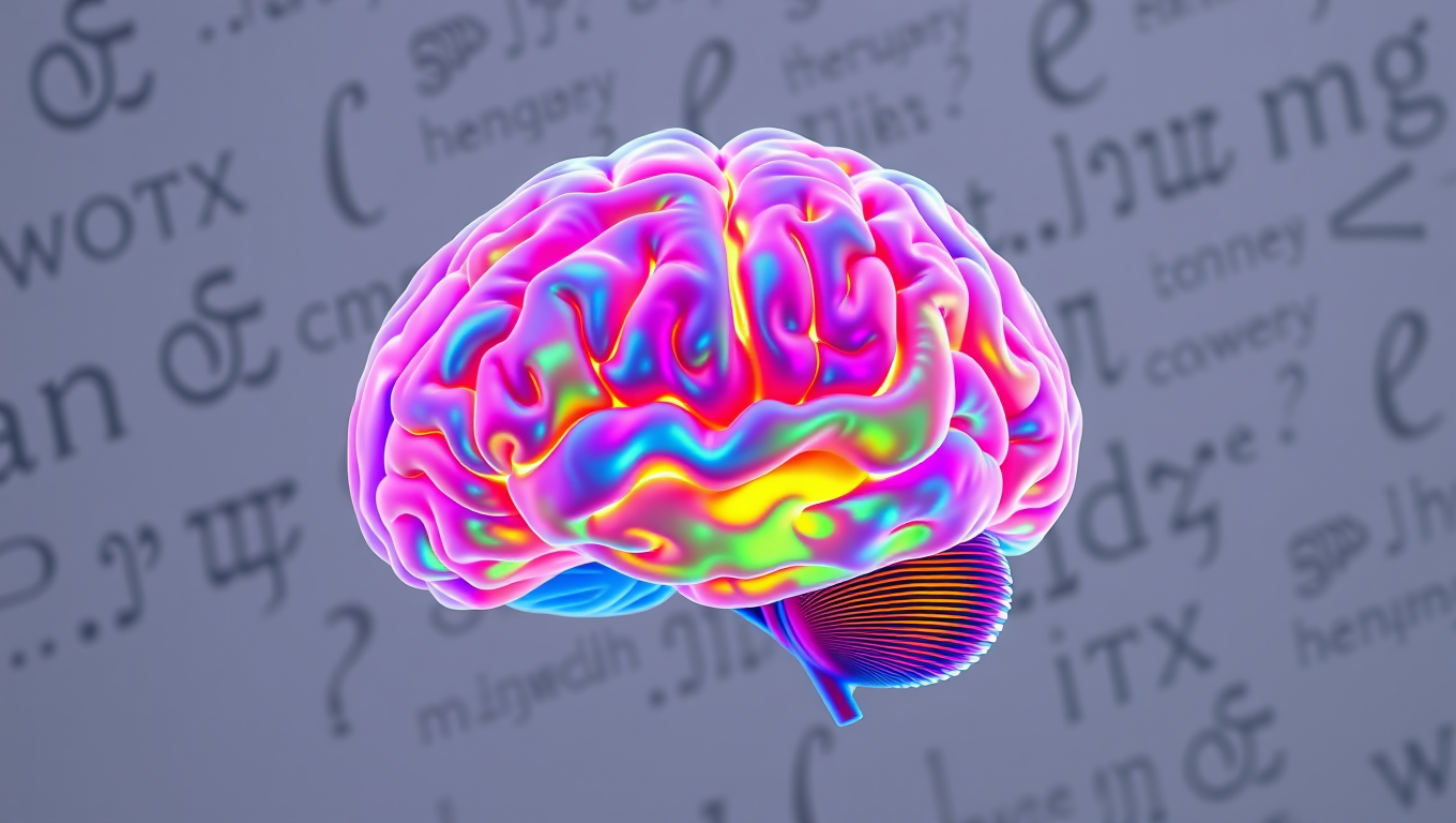
Researchers identified two brain networks involved in word retrieval — the cognitive process of accessing words we need to speak. A semantic network processes meaning in middle/inferior frontal gyri, while an articulatory network in inferior frontal/precentral gyri plans speech production.
Avatar photo

Published

on

The human brain is capable of performing incredible feats, from recalling memories to navigating complex mathematical equations. Yet, there lies one basic yet essential ability that often goes unnoticed – the power to name words we want to say. This seemingly simple act, called word retrieval, can be severely compromised in individuals with brain damage or neurological disorders. Despite decades of research, scientists have long sought to understand how the brain retrieves words during speech.

A groundbreaking study by researchers at New York University has shed light on this mystery, revealing a left-lateralized network in the dorsolateral prefrontal cortex that plays a crucial role in naming. Published in Cell Reports, the findings provide new insights into the neural architecture of language, with potential applications for both neuroscience and clinical interventions.

The study involved recording electrocorticographic (ECoG) data from 48 neurosurgical patients to examine the spatial and temporal organization of language processing in the brain. By using unsupervised clustering techniques, the researchers identified two distinct but overlapping networks responsible for word retrieval – a semantic processing network located in the middle and inferior frontal gyri, and an articulatory planning network situated in the inferior frontal and precentral gyri.

A striking ventral-dorsal gradient was observed in the prefrontal cortex, with articulatory planning localized ventrally and semantic processing uniquely represented in a dorsal region of the inferior frontal gyrus and middle frontal gyrus. This previously underappreciated hub for language processing has been found to play a crucial role in mapping sounds to meaning in an auditory context.

The findings have far-reaching implications, not only for theoretical neuroscience but also for clinical applications. Language deficits, such as anomia – the inability to retrieve words – are common in stroke, brain injury, and neurodegenerative disorders. Understanding the precise neural networks involved in word retrieval could lead to better diagnostics and targeted rehabilitation therapies for patients suffering from these conditions.

Additionally, the study provides a roadmap for future research in brain-computer interfaces (BCIs) and neuroprosthetics. By decoding the neural signals associated with naming, scientists could potentially develop assistive devices for individuals with speech impairments, allowing them to communicate more effectively through direct brain-computer communication.

In conclusion, our ability to name the world around us is not just a simple act of recall but the result of a sophisticated and finely tuned neural system – one that is now being revealed in greater detail than ever before. The discovery of this hidden brain network has opened up new avenues for research and potential applications, ultimately improving our understanding of human language and cognition.

Brain Injury

Unlocking the Secret to Weight Loss: Scientists Discover Key Brain Cells that Regulate Semaglutide’s Effects

A specific group of nerve cells in the brain stem appears to control how semaglutide affects appetite and weight — without causing nausea.

Avatar photo

Published

on

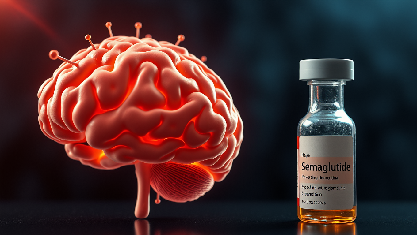
The discovery of a specific group of nerve cells in the brain stem has shed new light on how semaglutide affects appetite and weight loss. Researchers at the University of Gothenburg have made a groundbreaking find that could pave the way for better drugs to treat obesity.

Semaglutide, a GLP-1R agonist, is already well-established as part of the treatment for obesity and type 2 diabetes. However, it can cause side effects such as nausea and muscle loss. The researchers were able to distinguish the nerve cells in the brain that control the beneficial effects of semaglutide from those that contribute to side effects.

In a study published in Cell Metabolism, the researchers worked with mice and tracked which nerve cells were activated by semaglutide. They then stimulated these cells without administering the drug itself. The result was that the mice ate less and lost weight, just as they did when treated with semaglutide. When these nerve cells were killed, the drug’s effect on appetite and fat loss decreased significantly, but side effects such as nausea and muscle loss remained.

“This suggests that these nerve cells control the beneficial effects of semaglutide,” says Júlia Teixidor-Deulofeu, first author of the study. “We have therefore identified a specific group of nerve cells that is necessary for the effects that semaglutide has on weight and appetite, but which does not appear to contribute to any significant extent to side effects such as nausea.”

The identified nerve cells are located in an area of the brain called the dorsal vagal complex. The study provides new knowledge about how semaglutide works in the brain and deeper insight into how the brain stem regulates our energy balance.

“The better we understand this, the greater the opportunity we have to improve them,” says Linda Engström Ruud, researcher and supervisor to PhD students Júlia Teixidor-Deulofeu and Sebastian Blid Sköldheden, who both worked on the project.

This discovery has significant implications for the development of better drugs to treat obesity and could potentially lead to improved treatment options with fewer side effects.

Continue Reading

Brain Injury

Rapid Genetic Diagnosis of Brain Tumors Revolutionizes Care

Scientists and medics have developed an ultra-rapid method of genetically diagnosing brain tumors that will cut the time it takes to classify them from 6-8 weeks, to as little as two hours.The team utilized the new approach during 50 brain tumor surgeries to deliver rapid, intraoperative diagnoses. This approach has achieved a 100% success rate, providing diagnostic results in under two hours from surgery and detailed tumor classifications within minutes of sequencing.

Avatar photo

Published

on

The discovery of an ultra-rapid method for genetically diagnosing brain tumors has been hailed as a groundbreaking achievement by scientists and medics. This innovative approach can reduce the time it takes to classify brain tumors from 6-8 weeks to as little as two hours, providing quicker access to optimal care for thousands of patients each year in the UK.

The team at Nottingham University Hospitals NHS Trust (NUH) has successfully developed this method using portable sequencing devices and a software tool called ROBIN. This technology can quickly sequence specific parts of human DNA, allowing relevant information to be extracted and analyzed within minutes.

Brain tumors require complex genetic tests to diagnose, which traditionally takes weeks or even months to complete. The long wait for results is traumatic for patients and delays the start of radiotherapy and chemotherapy, potentially reducing the effectiveness of treatment.

The new method has achieved a 100% success rate in delivering rapid diagnoses during surgeries, providing accurate information within hours. This not only improves clinical decision-making but also reduces anxiety and worry for patients facing an already difficult time.

Experts believe that this technology will be a game-changer in diagnosing brain tumors, increasing the speed and accuracy of diagnoses while being more cost-effective than current methods. The team is now working to roll out this new testing across NHS Trusts in the UK, ensuring rapid access to optimal care for patients.

The potential impact on patient care is immense, with accurate diagnosis within hours of surgery transforming treatment options and removing uncertainty for patients. As one expert noted, “This technology will drive equity of access to rapid and accurate molecular diagnosis,” paving the way for personalized clinical trials and improved patient outcomes.

Continue Reading

Brain Injury

“Precision Gene Delivery to the Brain: A Breakthrough in Neuroscience Research”

Research teams funded by the National Institutes of Health (NIH) have created a versatile set of gene delivery systems that can reach different neural cell types in the human brain and spinal cord with exceptional accuracy. These delivery systems are a significant step toward future precise gene therapy to the brain that could safely control errant brain activity with high precision. In contrast, current therapies for brain disorders mostly treat only symptoms.

Avatar photo

Published

on

Scientists have made a groundbreaking discovery that could revolutionize our understanding of the human brain. A team of researchers funded by the National Institutes of Health (NIH) has developed a versatile set of gene delivery systems that can reach different neural cell types in the brain and spinal cord with exceptional accuracy.

This innovative platform has the potential to transform how scientists study neural circuits, providing them with gene delivery systems for various species used in research without the need for genetically modified animals. The new delivery tools use a small, stripped-down adeno-associated virus (AAV) to deliver DNA to target cells and can be broadly applied across many species and experimental systems.

The NIH’s Brain Research Through Advancing Innovative Neurotechnologies Initiative (BRAIN Initiative) has funded this large-scale project, which brings together experts in molecular biology, neuroscience, and artificial intelligence. The team has developed a comprehensive toolkit that includes standard operating procedures and user guides for these tools.

This collection of research tools will significantly accelerate understanding of the human brain. Importantly, the toolkit enables access to specific brain cell types in the prefrontal cortex, an area critical for decision-making and uniquely human traits. With other tools in the collection, scientists can better study individual cells and communication pathways known to be affected in several neurological diseases.

The new gene delivery systems lay the groundwork for more precise treatments that target only affected cells in the brain, spinal cord, or brain blood vessels. AAV-based treatments are already approved for some conditions, such as spinal muscular atrophy, which has transformed the lives of infants and young children who once faced severe disability or early death.

The toolkit is available at distribution centers like Addgene, a global supplier of genetic research tools. This collection of publications offers researchers standard operating procedures and user guides for these tools. The work is supported by the NIH’s BRAIN Initiative, which has provided funding to develop precise and reproducible access to cells and circuits in experimental research models of the brain and spinal cord.

Overall, this breakthrough in neuroscience research has the potential to revolutionize our understanding of the human brain and pave the way for more effective treatments for neurological diseases.

Continue Reading

Trending