Connect with us

Biochemistry Research

Unzipping DNA: Scientists Capture Molecular Movie Revealing Atomic-Level Process

Scientists have captured the first detailed ‘molecular movie’ showing DNA being unzipped at the atomic level — revealing how cells begin the crucial process of copying their genetic material.
Avatar photo

Published

on

Unzipping DNA: Scientists Capture Molecular Movie Revealing Atomic-Level Process

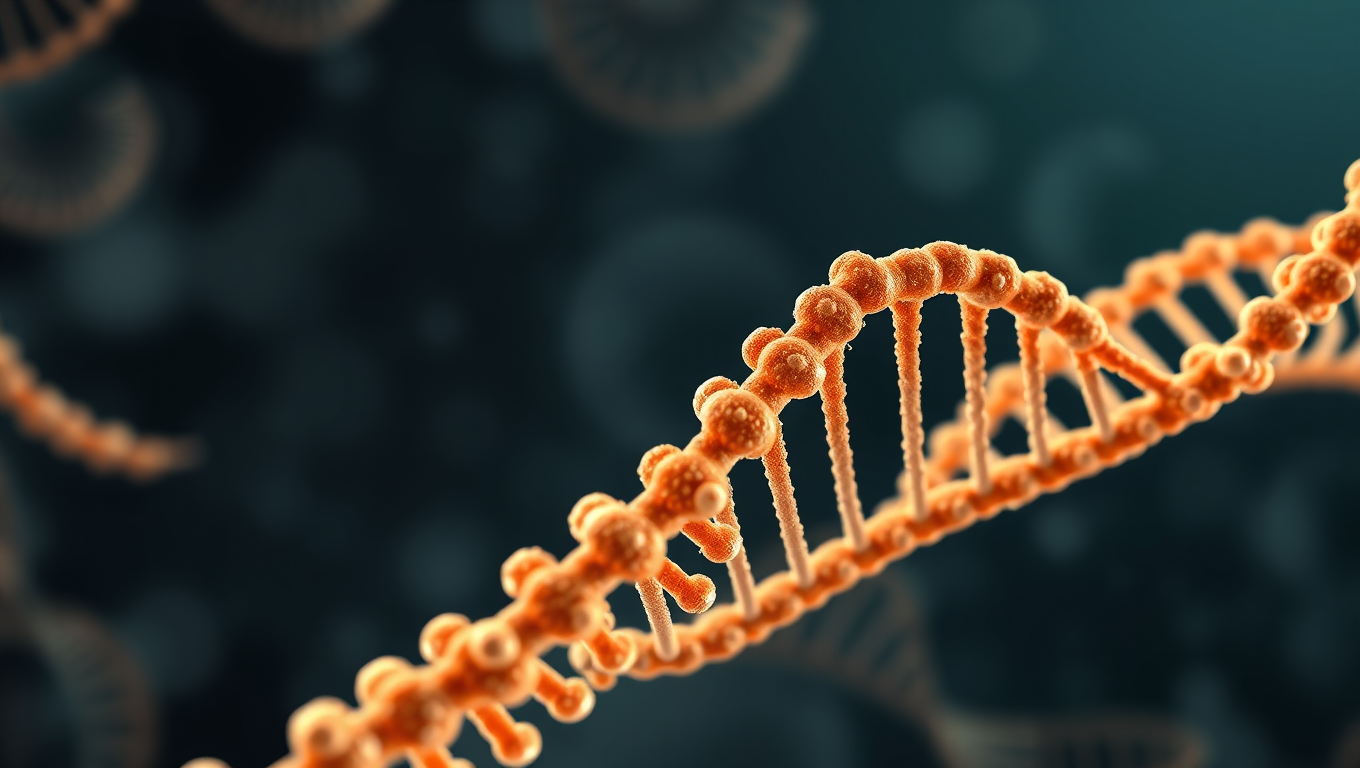
Scientists at the University of Leicester have made a groundbreaking discovery by capturing the first detailed molecular movie showing DNA being unzipped at the atomic level. This breakthrough revelation could have far-reaching implications for understanding how certain viruses and cancers replicate.

Using cutting-edge cryo-electron microscopy, the team was able to visualize a helicase enzyme (nature’s DNA unzipping machine) in the process of unwinding DNA. Helicases are essential during DNA replication as they separate double-stranded DNA into single strands, allowing each strand to be copied.

Dr. Taha Shahid, lead author on the paper and researcher at the University of Leicester’s Institute of Structural and Chemical Biology, explained: “We recorded multiple snapshots showing how this molecular motor methodically separates the DNA double helix. It’s like a molecular-scale zipper in action.”

The researchers found that rather than working by brute force as previously assumed, the helicase operates through an elegant mechanism that uses cellular fuel (ATP) as a precise trigger. The enzyme functions like a six-piston molecular engine, where each piston “fires” in sequence, driving the machine forward along the DNA.

Crucially, instead of pushing the strands apart directly, it releases built-up tension – like letting go of a compressed spring – allowing the DNA to unwind naturally. This “entropy switch” mechanism is fundamentally different from how scientists thought molecular motors worked.

The research was an international collaboration between the University of Leicester and King Abdullah University of Science and Technology (KAUST) in Saudi Arabia, which provided core funding. The Midlands Regional Cryo-EM Facility at the Leicester Institute of Structural and Chemical Biology also provided essential infrastructure for the work.

Dr. Shahid continued: “The helicase mechanism we’ve uncovered appears to be evolutionarily conserved from viruses to humans, providing a universal blueprint for understanding DNA replication across all domains of life.”

This discovery could guide the development of precisely targeted antiviral therapies that disrupt viral replication without harming human cells. Furthermore, the detailed structural insights gained from this research may inspire the design of synthetic molecular devices using similar principles.

Professor John Schwabe, Director of Leicester’s Institute for Structural and Chemical Biology, added: “This is another example of how our world-leading facility is contributing to revealing the critical fundamental mechanisms that underpin life.”

Bacteria

Unlocking Efficiency: Researchers Reveal Secrets of Cell Division with Min Proteins

The Min protein system prevents abnormal cell division in bacteria, but is poorly understood. Researchers have uncovered how engineered e.coli bacteria control protein levels for maximum efficiency.

Avatar photo

Published

on

The Min protein system is a complex process that helps bacteria divide evenly and correctly. For decades, scientists have studied this system, but understanding how it works efficiently has been a challenge. Recently, researchers at the University of California San Diego (UCSD) made a groundbreaking discovery that sheds new light on the efficiency of cell division.

The UCSD team developed a way to control Min protein expression levels independently in E. coli cells. This allowed them to observe how different concentrations of Min proteins affect the oscillations between the poles of the cell. The results were surprising: despite varying concentrations, the oscillations remained stable across a wide range, with E. coli producing just the right amount of Min proteins.

This breakthrough is significant because it shows that the Min protein system can efficiently guide division to the correct location without relying on precise control over protein levels. This finding has far-reaching implications for our understanding of cellular organization and function.

The study was published in Nature Physics, a leading scientific journal, and was funded by the National Institutes of Health (NIH). The research team consisted of experts from both physics and chemistry/biochemistry departments at UCSD, highlighting the importance of interdisciplinary collaboration in advancing our knowledge of cellular biology.

Continue Reading

Biochemistry Research

Unlocking Cell Movement: Researchers Crack the Code on How Cells Travel Through the Body

Scientists have discovered how chemokines and G protein-coupled receptors selectively bind each other to control how cells move.

Avatar photo

Published

on

Researchers from St. Jude Children’s Research Hospital and the Medical College of Wisconsin have made a groundbreaking discovery that sheds light on how cells travel through the body. By developing a data science framework, they were able to analyze chemokines and their associated G protein-coupled receptors (GPCRs), which are proteins that govern cell movement.

The scientists found that specific positions within structured and disordered regions of both proteins determine how chemokines and GPCRs bind each other. This understanding enabled them to artificially change chemokine-GPCR binding preferences and alter the resulting cell migration. Their findings have significant implications for disease treatment, such as enhancing cellular therapies’ ability to reach tumor sites, and increasing clarity about healthy processes like heart and blood vessel development.

Cell migration is a crucial process that influences many aspects of our bodies, including how immune cells travel to infection sites, brain development, and wound repair. However, the vast similarities between members of each protein family have presented a challenge in understanding how correct pairs form and control cell movement. The researchers’ data-driven approach identified the exact parts of each protein governing their molecular interactions.

“We found that cells have an elegant system that uses structure and disorder together to control cell migration,” said senior co-corresponding author M. Madan Babu, PhD. “With this understanding, we can now rationally introduce small changes in a chemokine’s structure to ultimately alter cell migration in desired ways.”

The scientists compared all human chemokine-binding GPCRs and all chemokines, then compared similar chemokines and GPCRs from other species. They also looked at each protein individually at a population level, finding places that stayed the same across groups and those that differed.

“Through our data analysis, we discovered that the information for how chemokines and GPCRs select for each other is stored in small, discrete packages of highly unstructured, disordered regions,” said first and co-corresponding author Andrew Kleist, MD. “The mix of those small packages from both the chemokine and receptor results in the unique interaction, similar to website data encryption keys, which governs cell migration.”

This discovery has significant implications for disease treatment and therapy development. The researchers’ framework can guide exploration into new medicines and improvements for existing cellular therapies.

“Now that we’ve shown a proof of concept, our approach will guide exploration into new medicines and improvements for existing cellular therapies,” Kleist said. “For example, it may be possible to create molecules that better lead immune cells to cancers or help recruit more blood stem cells for bone marrow transplants.”

The framework is freely available online at: https://github.com/andrewbkleist/chemokine_gpcr_encoding.

When people think about the body, we often think every cell stays in place. However, that’s a simplistic view. Depending on the tissue, cells are moving all the time, and our new understanding of those systems opens novel avenues for therapeutic development.

This discovery has the potential to revolutionize our understanding of cell movement and its role in various biological processes. By unlocking the code of cell movement, researchers can develop more effective treatments and therapies that target specific aspects of cellular behavior.

Continue Reading

Biochemistry

“Tailoring Gene Editing with Machine Learning: A Breakthrough in CRISPR-Cas9 Enzyme Engineering”

Genome editing has advanced at a rapid pace with promising results for treating genetic conditions — but there is always room for improvement. A new paper showcases the power of scalable protein engineering combined with machine learning to boost progress in the field of gene and cell therapy. In their study, authors developed a machine learning algorithm — known as PAMmla — that can predict the properties of about 64 million genome editing enzymes. The work could help reduce off-target effects and improve editing safety, enhance editing efficiency, and enable researchers to predict customized enzymes for new therapeutic targets.

Avatar photo

Published

on

The article “Tailoring Gene Editing with Machine Learning: A Breakthrough in CRISPR-Cas9 Enzyme Engineering” discusses how researchers from Mass General Brigham have harnessed machine learning to revolutionize the field of genome editing. By developing a machine learning algorithm called PAMmla, they’ve predicted the properties of over 64 million genome editing enzymes, significantly expanding our repertoire of effective and safe CRISPR-Cas9 enzymes.

CRISPR-Cas9 enzymes are powerful tools for editing genes, but their traditional application can have off-target effects, modifying DNA at unintended sites in the genome. The researchers’ novel approach uses machine learning to better predict and tailor these enzymes, ensuring greater specificity and accuracy in gene editing. This scalable solution has the potential to transform our understanding of genetic conditions and unlock new therapeutic targets.

The study showcases the power of PAMmla by demonstrating its utility in precise editing disease-causing sequences in primary human cells and mice. The researchers have also made a web tool available for others to use this model, enabling the community to create customized enzymes tailored for specific research and therapeutic applications.

Ben Kleinstiver, PhD, and Rachel A. Silverstein, PhD candidate, are leading authors on this study, highlighting the potential of machine learning in expanding our capabilities in gene editing. This breakthrough has significant implications for the field, offering a new era of precision and safety in genome editing technology.

Continue Reading

Trending