Connect with us

Birth Defects

Pregnancy Triggers Lasting Changes to the Mouse Intestine

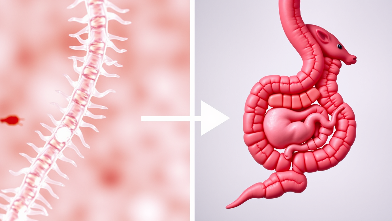
Researchers have found that the small intestine grows in response to pregnancy in mice. This partially irreversible change may help mice support a pregnancy and prepare for a second.
Avatar photo

Published

on

The Francis Crick Institute has made a groundbreaking discovery: the small intestine grows in response to pregnancy in mice. This irreversible change may help mice support a pregnancy and prepare for a second. The research team, led by Irene Miguel-Aliaga, found that pregnant mice had a longer small intestine from just seven days into the pregnancy. By the end of the pregnancy, around day 18, the small intestine was 18% longer.

Interestingly, the small intestine remained longer after birth, even up to 35 days after the mice had finished lactating. This suggests that the change isn’t reversible and might be a trade-off in energy. The team observed that the small intestine was even longer after a second pregnancy than the first.

Inside the small intestine, the villi (protrusions that absorb nutrients) and crypts (where the cells supplying the villi are produced) became longer and deeper at the same time as the gut lengthened. However, both returned to pre-pregnancy values just seven days after weaning.

The researchers altered the diet of the mice, including probiotic intake, to show that these physiological changes in pregnancy occurred regardless of nutritional change. They then took a deeper dive into what was happening in the villi.

They found that precursors of intestinal epithelial cells were rapidly growing in number and newly generated cells migrated faster up the villi in early pregnancy. These effects continued after birth into lactation but returned to pre-pregnancy rates just seven days after weaning.

By looking at which genes were switched on during pregnancy, the researchers saw the biggest number of genetic changes in enterocytes, nutrient-absorbing cells in the villi that are produced by precursor cells in the crypt. These changes were largely related to increasing metabolic activity.

One of the metabolic effects observed at a very early stage was an increase in a membrane protein called SGLT3a. Unlike other SGLT proteins, SGLT3a does not sense glucose levels outside of the cell but responds to sodium and protons instead. An increase in SGLT3a was responsible for about 45% of the villi growth triggered by reproduction, including cell expansion, but wasn’t necessary for entire small intestine lengthening.

The team believes that hormones triggered by reproduction may play a role in switching on the gene for SGLT3a, as pseudo-pregnant mice – females whose pregnancy hormone levels have increased after mating with sterile males – still showed some villi growth and intestinal lengthening.

Irene Miguel-Aliaga said: “We don’t often think about organs changing size or appearance in response to triggers in adulthood rather than earlier childhood and adolescent development, but the gut is a striking example of how the body responds to a new challenge at different stages of life, in this case pregnancy.”

Tomotsune Ameku, former postdoctoral researcher at the Crick, now Assistant Professor at Science Tokyo and first author of the study, said: “Understanding how pregnancy impacts the body in other mammals is a critical first step to understanding this in people. We don’t fully understand why the gut expands in response to pregnancy, but we think it must give an evolutionary advantage and help mice reproduce regularly.”

The researchers are now investigating whether other cell types in the mouse intestine also experience remodelling during pregnancy, and whether this happens in humans, by looking at gut length in people who have and haven’t had children.

Birth Defects

“Breakthrough in Down Syndrome Research: 40Hz Light and Sound Stimulation Improves Cognition and Neurogenesis”

A new study provides new evidence that sensory stimulation of a gamma-frequency brain rhythm may promote broad-based restorative neurological health response.

Avatar photo

Published

on

The study, conducted at The Picower Institute for Learning and Memory and Alana Down Syndrome Center at MIT, found that exposing mice genetically engineered to model Down syndrome to 40Hz light and sound stimulation improved cognition, circuit connectivity, and encouraged the growth of new neurons. This breakthrough has significant implications for individuals with Down syndrome, who often experience cognitive impairments.

The research team, led by Md Rezaul Islam and Brennan Jackson, used a technique called single cell RNA sequencing to examine gene expression in individual neurons. They found that many genes related to forming and organizing neural circuit connections were directly affected by the 40Hz stimulation. Furthermore, they observed an increase in synapses in a critical subregion of the hippocampus.

The study also showed that the 40Hz-stimulated mice exhibited more than unstimulated mice in neurogenesis, which is essential for generating new neurons. The researchers hypothesized that this increase in new neurons likely helps explain at least some of the increase in new synapses and improved short-term memory function.

This breakthrough has significant implications for individuals with Down syndrome, who often experience cognitive impairments. It also adds evidence that 40Hz-stimulated mice mount a homeostatic response to aberrations caused by disease pathology, be it neurodegeneration in Alzheimer’s or deficits of neurogenesis in Down syndrome.

However, the authors cautioned that the study had limits, including the use of an imperfect mouse model and male mice only. They also emphasized the need for further research, particularly examining changes in other cognitively critical brain regions such as the prefrontal cortex.

Continue Reading

Birth Defects

Breaking Down Barriers: Towards Gene-Targeting Drugs for Brain Diseases

Getting therapeutic drugs past the blood-brain barrier has long been a major challenge in treating brain diseases. Now, researchers have explored how cholesterol-modified heteroduplex oligonucleotides (Chol-HDOs) enhance drug delivery to the brain. Their study reveals that Chol-HDOs bind tightly to serum proteins, allowing them to persist in the bloodstream and cross into brain tissue. These findings offer insights into gene-targeting therapies and could help develop treatments for conditions like Alzheimer’s disease.

Avatar photo

Published

on

Breaking Down Barriers: Towards Gene-Targeting Drugs for Brain Diseases

The human brain is a complex and intricate organ that has long been a challenge to treat when it comes to diseases like Alzheimer’s, Parkinson’s, and brain cancers. One of the major obstacles in delivering therapeutic drugs to the brain is the blood-brain barrier (BBB), a protective layer that restricts the passage of molecules from the bloodstream into the brain.

To overcome this hurdle, researchers at Tokyo University of Science have been exploring new ways to deliver gene-targeting drugs, specifically antisense oligonucleotides (ASOs) and heteroduplex oligonucleotides (HDOs), directly to the brain. In a recent study published in the Journal of Controlled Release, the team led by Professor Makiya Nishikawa demonstrated that modifying HDOs with cholesterol molecules (Chol-HDOs) could improve their stability and specificity, allowing them to penetrate the cerebral cortex beyond the blood vessels.

The key to this success lies in how Chol-HDOs interact with proteins in the bloodstream. Unlike ASOs and HDOs, which bind electrostatically to serum proteins with low affinity and are taken up by cells, Chol-HDOs bind tightly to serum proteins, including lipoproteins, via hydrophobic interactions. This strong binding results in slow clearance from the bloodstream, allowing Chol-HDOs to remain in circulation for a longer period.

The researchers also showed that inhibiting scavenger receptors in cells reduces the uptake of both ASOs and Chol-HDOs in the liver and kidneys, shedding light on how these compounds are taken up by different organs. This finding has significant implications for the design of brain-targeting drugs based on Chol-HDOs.

With over 55 million people living with dementia worldwide and 300,000 cases of brain cancer reported annually, the potential therapeutic applications of modified HDOs are vast. The possibility of efficiently delivering ASOs and other nucleic acid-based drugs to the brain may lead to the development of treatments for brain diseases with significant unmet medical needs.

This study provides valuable insight into how brain-targeting drugs could be designed based on Chol-HDOs, paving the way for a new generation of compounds that effectively target brain diseases. As research continues, we can expect modified HDOs to offer hope to millions of patients and their families around the world.

Continue Reading

Birth Defects

Severe Heart Rhythm Disorders Persist After Valve Surgery in Some Patients

People with a certain heart valve abnormality are at increased risk of severe heart rhythm disorders, even after successful valve surgery. The condition is more common in women and younger patients with valve disorder and can, in the worst case, lead to sudden cardiac arrest.

Avatar photo

Published

on

Severe Heart Rhythm Disorders Persist After Valve Surgery in Some Patients

A recent study from Karolinska Institutet and Karolinska University Hospital in Sweden has found that some patients who undergo heart surgery for valve disorders may still be at risk of severe heart rhythm disorders, even after a successful operation. The study, published in the European Heart Journal, highlights the importance of closely monitoring these patients to prevent cardiac arrest.

The condition being studied is called mitral annular disjunction (MAD), which affects the mitral valve attachment and can lead to severe arrhythmias. Researchers at Karolinska Institutet investigated 599 patients with mitral valve prolapse who underwent heart surgery between 2010 and 2022, and found that 16% of them also had MAD.

The study showed that people with MAD were more likely to be female and younger than those without the condition, and had more extensive mitral valve disease. Although the surgery was successful in correcting MAD, these patients still had a higher risk of ventricular arrhythmias during five years of follow-up compared to patients without preoperative MAD.

“This is a concerning finding that highlights the need for closer monitoring of these patients after surgery,” said Bahira Shahim, associate professor at the Department of Medicine, Solna, Karolinska Institutet and cardiologist at Karolinska University Hospital. “Our results show that it’s essential to keep an eye on these patients even after a successful operation.”

The researchers are now investigating several hypotheses, including whether MAD causes permanent changes in the heart muscle over time or is a sign of underlying heart muscle disease. They are using MRI and analyzing tissue samples from the heart muscle to further study the condition.

The research was led by cardiologist and associate professor Bahira Shahim, along with Magnus Dalén and Klara Lodin. It was financed by ALF funds, the Swedish Heart-Lung Foundation, the Swedish Research Council, the Swedish Society for Medical Research, the Swedish Society of Medicine, and Karolinska Institutet.

In conclusion, this study highlights the importance of monitoring patients who have undergone valve surgery to prevent severe heart rhythm disorders, even after a successful operation. Further research is needed to fully understand the mechanisms behind MAD and how to best treat it.

Continue Reading

Trending