
While we try to keep things accurate, this content is part of an ongoing experiment and may not always be reliable.
Please double-check important details — we’re not responsible for how the information is used.
Disorders and Syndromes
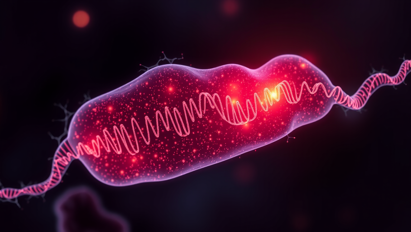
“Placenta Plays Key Role in Genetic Risk for Schizophrenia and Other Neuropsychiatric Disorders”
An international team has identified associations between modifications in the placenta and the risk of developing schizophrenia, bipolar disorder, and major depression disorder.


Alzheimer's
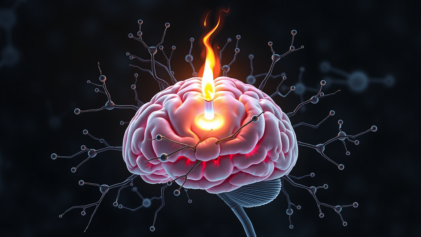
Scientists Unlock Secret to Reversing Memory Loss by Boosting Brain’s Energy Engines
Scientists have discovered a direct cause-and-effect link between faulty mitochondria and the memory loss seen in neurodegenerative diseases. By creating a novel tool to boost mitochondrial activity in mouse models, researchers restored memory performance, suggesting mitochondria could be a powerful new target for treatments. The findings not only shed light on the early drivers of brain cell degeneration but also open possibilities for slowing or even preventing diseases like Alzheimer’s.
Anger Management
The Hidden Depression Crisis in Early Menopause: Uncovering the Unexpected Risks
Premature menopause isn t just a hormonal issue it s a deeply emotional one for many women. A new study reveals that almost 30% experience depression, and it s not just about hormone loss but also grief, identity, and support systems.

Alternative Medicine
“The Sleeping Giants: How Tai Chi, Yoga, and Jogging Rival Pills for Beating Insomnia”
Yoga, Tai Chi, walking, and jogging may be some of the best natural remedies for improving sleep and tackling insomnia, according to a large analysis comparing various treatments. While cognitive behavioral therapy (CBT) remains effective, exercise-based approaches—especially Tai Chi—were shown to deliver significant improvements in total sleep time, efficiency, and reducing how long people stay awake after falling asleep. Yoga stood out for boosting overall restfulness, and jogging helped ease insomnia symptoms.


A New Horizon for Vision: How Gold Nanoparticles May Restore People’s Sight

Revolutionizing Quantum Communication: Direct Connections Between Multiple Processors

Retiring Abroad Can Be Lonely Business
Harnessing Water Waves: A Breakthrough in Controlling Floating Objects

“Unveiling Hidden Patterns: A New Twist on Interference Phenomena”

Household Electricity Three Times More Expensive Than Upcoming ‘Eco-Friendly’ Aviation E-Fuels, Study Reveals
Reducing Falls Among Elderly Women with Polypharmacy through Exercise Intervention
-
 Detectors1 year ago
A New Horizon for Vision: How Gold Nanoparticles May Restore People’s Sight
-
Cancer1 year ago
Revolutionizing Quantum Communication: Direct Connections Between Multiple Processors
-
Earth & Climate1 year ago
Retiring Abroad Can Be Lonely Business
-
Albert Einstein1 year ago
Harnessing Water Waves: A Breakthrough in Controlling Floating Objects
-
Chemistry1 year ago
“Unveiling Hidden Patterns: A New Twist on Interference Phenomena”
-
Earth & Climate1 year ago
Household Electricity Three Times More Expensive Than Upcoming ‘Eco-Friendly’ Aviation E-Fuels, Study Reveals
-
Diseases and Conditions1 year ago
Reducing Falls Among Elderly Women with Polypharmacy through Exercise Intervention
-
Agriculture and Food1 year ago
“A Sustainable Solution: Researchers Create Hybrid Cheese with 25% Pea Protein”


